Selection and structure of ion-selective ligands for platelet integrin alpha IIb(beta) 3.
Smith, J.W., Le Calvez, H., Parra-Gessert, L., Preece, N.E., Jia, X., Assa-Munt, N.(2002) J Biol Chem 277: 10298-10305
- PubMed: 11748219
- DOI: https://doi.org/10.1074/jbc.M108071200
- Primary Citation of Related Structures:
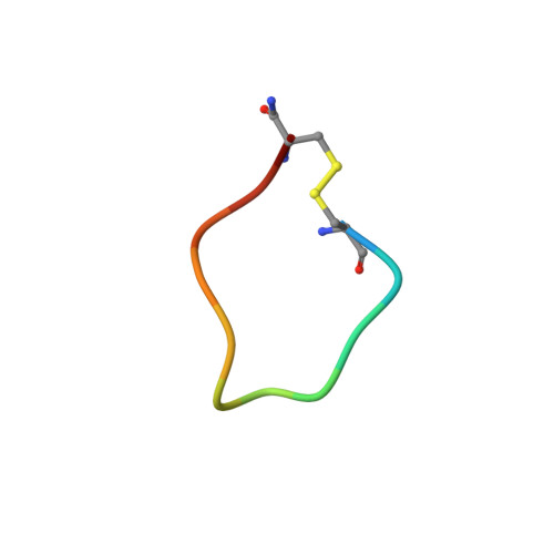
1I6Y, 1I8E, 1I93, 1I98 - PubMed Abstract:
Integrins contain a number of divalent cation binding sites that control ligand binding affinity. Ions such as Ca(2+) and Mg(2+) bind to distinct sites on integrin and can have opposing effects on ligand binding. These effects are presumably brought about by alterations of the shape of the ligand binding pocket. To gain insight into the nature of these structural differences, we probed the integrin ligand binding site with an RGD-based library of unparalleled complexity. A cysteine-constrained phage library containing six random amino acids and the RGD motif present in seven different registers was used to select for ligands that exhibit ion-selective binding to integrin alpha(IIb)beta(3). The library was used to select for peptides that bind to the integrin alpha(IIb)beta(3) preferentially in Ca(2+) versus Mg(2+). Peptides were identified which bound selectively in each ion. The Ca(2+)-selective peptides had a range of sequences, with the only obvious consensus involving a motif that had four cysteine residues bonded in a 1,4:2,3 arrangement. Interestingly though, the Mg(2+)-selective peptides exhibited a well defined consensus motif containing Cys-X-aromatic-L/G-R-G-D-hydrophobic-R-R/K-Cys. As a first step toward understanding the structural basis for this selectivity, solution NMR structures were obtained for representatives of both sets of peptides. All peptides formed turns, with the RGD motif at the apex. The Mg(2+)-selected peptides contained a unique basic patch that protrudes from the base of the turn.
Organizational Affiliation:
Program on Cell Adhesion, Cancer Research Center, the Burnham Institute, La Jolla, California 92037, USA. jsmith@burnham.org