Human interleukin-4 and variant R88Q: phasing X-ray diffraction data by molecular replacement using X-ray and nuclear magnetic resonance models.
Muller, T., Oehlenschlager, F., Buehner, M.(1995) J Mol Biol 247: 360-372
- PubMed: 7707380
- DOI: https://doi.org/10.1006/jmbi.1994.0144
- Primary Citation of Related Structures:
1HIJ, 1HIK - PubMed Abstract:
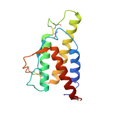
The structure of recombinant human interleukin-4 (hIL-4) has been determined by both NMR and X-ray diffraction methods in several laboratories, including ours. The X-ray and NMR structures were successfully applied for solving the X-ray crystal structure by molecular replacement. Due to the small size of the hIL-4 molecule (129 residues) and its lack of structural diversity (4-helix bundle), this task was especially difficult and required special care with rotation function applications. The crucial point was that proper removal of the Patterson origin peaks was indispensable in all cases. All available structures of hIL-4 were checked, in a standardized procedure, for their suitability as templates for molecular replacement. The models derived from the various structures are close to, but not in all loop details identical with, the genuine X-ray structures. The deviations of the X-ray structure-derived models are of the same magnitude as the differences between the original X-ray structures, while the deviations of the NMR structure-derived models are two to three times as large. The hIL-4 variant R88Q is a binding mutant, its affinity to the receptor is decreased by a factor of about 200. Its X-ray structure was determined by molecular replacement using the wild-type X-ray structure determined in our laboratory as a model. The structure of R88Q is virtually identical with that of the wild-type protein. All differences besides the shortened side-chain of residue 88 occur at surface residues with high temperature factors, i.e. at spots where the structure is not well defined. Since the structure is not perturbed, the biological effect of decreased receptor affinity has to be attributed to the loss of a single positive charge in the surface area of the main receptor contact.
Organizational Affiliation:
Physiologische Chemie, Theodor-Boveri-Institut für Biowissenschaften, (Biozentrum) der Universität Würzburg, Germany.