1HG2

CALM-N N-terminal domain of clathrin assembly lymphoid myeloid leukaemia protein, Inositol(4,5)P2 complex


Experimental Data Snapshot

  • Method: X-RAY DIFFRACTION
  • Resolution: 2.00 Å
  • R-Value Free: 0.217 
  • R-Value Work: 0.197 
  • R-Value Observed: 0.198 

wwPDB Validation   3D Report Full Report


Ligand Structure Quality Assessment 


This is version 1.4 of the entry. See complete history


Literature

Simultaneous Binding of Ptdins(4,5)P2 and Clathrin by Ap180 in the Nucleation of Clathrin Lattices on Membranes

Ford, M.G.J.Pearse, B.M.F.Higgins, M.K.Vallis, Y.Owen, D.J.Gibson, A.Hopkins, C.R.Evans, P.R.Mcmahon, H.T.

(2001) Science 291: 1051

  • DOI: https://doi.org/10.1126/science.291.5506.1051
  • Primary Citation of Related Structures:  
    1HF8, 1HFA, 1HG2, 1HG5

  • PubMed Abstract: 

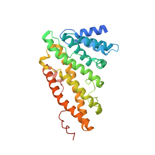
    Adaptor protein 180 (AP180) and its homolog, clathrin assembly lymphoid myeloid leukemia protein (CALM), are closely related proteins that play important roles in clathrin-mediated endocytosis. Here, we present the structure of the NH2-terminal domain of CALM bound to phosphatidylinositol-4,5- bisphosphate [PtdIns(4,5)P2] via a lysine-rich motif. This motif is found in other proteins predicted to have domains of similar structure (for example, Huntingtin interacting protein 1). The structure is in part similar to the epsin NH2-terminal (ENTH) domain, but epsin lacks the PtdIns(4,5)P2-binding site. Because AP180 could bind to PtdIns(4,5)P2 and clathrin simultaneously, it may serve to tether clathrin to the membrane. This was shown by using purified components and a budding assay on preformed lipid monolayers. In the presence of AP180, clathrin lattices formed on the monolayer. When AP2 was also present, coated pits were formed.


  • Organizational Affiliation

    Medical Research Council (MRC) Laboratory of Molecular Biology, Hills Road, Cambridge, CB2 2QH, UK.


Macromolecules
Find similar proteins by:  (by identity cutoff)  |  3D Structure
Entity ID: 1
MoleculeChains Sequence LengthOrganismDetailsImage
CLATHRIN ASSEMBLY PROTEIN SHORT FORM289Rattus norvegicusMutation(s): 0 
UniProt
Find proteins for O55012 (Rattus norvegicus)
Explore O55012 
Go to UniProtKB:  O55012
Entity Groups  
Sequence Clusters30% Identity50% Identity70% Identity90% Identity95% Identity100% Identity
UniProt GroupO55012
Sequence Annotations
Expand
  • Reference Sequence
Small Molecules
Ligands 1 Unique
IDChains Name / Formula / InChI Key2D Diagram3D Interactions
IP2
Query on IP2

Download Ideal Coordinates CCD File 
B [auth A]D-MYO-INOSITOL-4,5-BISPHOSPHATE
C6 H14 O12 P2
MCKAJXMRULSUKI-UZAAGFTCSA-N
Experimental Data & Validation

Experimental Data

  • Method: X-RAY DIFFRACTION
  • Resolution: 2.00 Å
  • R-Value Free: 0.217 
  • R-Value Work: 0.197 
  • R-Value Observed: 0.198 
  • Space Group: P 41 21 2
Unit Cell:
Length ( Å )Angle ( ˚ )
a = 77.859α = 90
b = 77.859β = 90
c = 122.333γ = 90
Software Package:
Software NamePurpose
REFMACrefinement
MOSFLMdata reduction
SCALAdata scaling

Structure Validation

View Full Validation Report


Ligand Structure Quality Assessment 


Entry History 

Deposition Data

Revision History  (Full details and data files)

  • Version 1.0: 2001-02-12
    Type: Initial release
  • Version 1.1: 2011-05-08
    Changes: Version format compliance
  • Version 1.2: 2011-07-13
    Changes: Version format compliance
  • Version 1.3: 2019-10-09
    Changes: Data collection, Experimental preparation, Other
  • Version 1.4: 2023-12-13
    Changes: Data collection, Database references, Refinement description