1H97

Trematode hemoglobin from Paramphistomum epiclitum


Experimental Data Snapshot

  • Method: X-RAY DIFFRACTION
  • Resolution: 1.17 Å
  • R-Value Free: 0.173 
  • R-Value Observed: 0.121 

wwPDB Validation   3D Report Full Report


This is version 1.4 of the entry. See complete history


Literature

Very High Resolution Structure of a Trematode Hemoglobin Displaying a Tyrb10-Tyre7 Heme Distal Residue Pair and High Oxygen Affinity

Pesce, A.Dewilde, S.Kiger, L.Milani, M.Ascenzi, P.Marden, M.C.Van, M.L.Vanfleteren, J.Moens, L.Bolognesi, M.

(2001) J Mol Biol 309: 1153

  • DOI: https://doi.org/10.1006/jmbi.2001.4731
  • Primary Citation of Related Structures:  
    1H97

  • PubMed Abstract: 

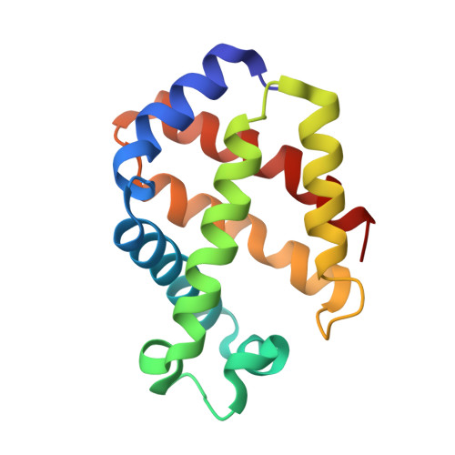
    Monomeric hemoglobin from the trematode Paramphistomum epiclitum displays very high oxygen affinity (P(50)<0.001 mm Hg) and an unusual heme distal site containing tyrosyl residues at the B10 and E7 positions. The crystal structure of aquo-met P. epiclitum hemoglobin, solved at 1.17 A resolution via multiwavelength anomalous dispersion techniques (R-factor=0.121), shows that the heme distal site pocket residue TyrB10 is engaged in hydrogen bonding to the iron-bound ligand. By contrast, residue TyrE7 is unexpectedly locked next to the CD globin region, in a conformation unsuitable for heme-bound ligand stabilisation. Such structural organization of the E7 distal residue differs strikingly from that observed in the nematode Ascaris suum hemoglobin (bearing TyrB10 and GlnE7 residues), which also displays very high oxygen affinity. The oxygenation and carbonylation parameters of wild-type P. epiclitum Hb as well as of single- and double-site mutants, with residue substitutions at positions B10, E7 and E11, have been determined and are discussed here in the light of the protein atomic resolution crystal structure.


  • Organizational Affiliation

    Department of Physics-INFM, Advanced Biotechnology Centre, University of Genova, Largo Rosanna Benzi, 10, Genova, I-16132, Italy.


Macromolecules
Find similar proteins by:  (by identity cutoff)  |  3D Structure
Entity ID: 1
MoleculeChains Sequence LengthOrganismDetailsImage
Globin-3
A, B
147Paramphistomum epiclitumMutation(s): 0 
UniProt
Find proteins for P80721 (Paramphistomum epiclitum)
Explore P80721 
Go to UniProtKB:  P80721
Entity Groups  
Sequence Clusters30% Identity50% Identity70% Identity90% Identity95% Identity100% Identity
UniProt GroupP80721
Sequence Annotations
Expand
  • Reference Sequence
Experimental Data & Validation

Experimental Data

  • Method: X-RAY DIFFRACTION
  • Resolution: 1.17 Å
  • R-Value Free: 0.173 
  • R-Value Observed: 0.121 
  • Space Group: P 21 21 21
Unit Cell:
Length ( Å )Angle ( ˚ )
a = 38.7α = 90
b = 83.7β = 90
c = 94.6γ = 90
Software Package:
Software NamePurpose
SHELXL-97refinement
DENZOdata reduction
SCALEPACKdata scaling
SOLVEphasing

Structure Validation

View Full Validation Report


Entry History 

Deposition Data

Revision History  (Full details and data files)

  • Version 1.0: 2001-06-21
    Type: Initial release
  • Version 1.1: 2012-09-19
    Changes: Advisory, Atomic model, Data collection, Database references, Derived calculations, Non-polymer description, Other, Refinement description, Structure summary, Version format compliance
  • Version 1.2: 2013-12-04
    Changes: Source and taxonomy
  • Version 1.3: 2019-07-24
    Changes: Advisory, Data collection
  • Version 1.4: 2019-10-23
    Changes: Data collection, Database references, Other, Source and taxonomy, Structure summary