1H7O

SCHIFF-BASE COMPLEX OF YEAST 5-AMINOLAEVULINIC ACID DEHYDRATASE WITH 5-AMINOLAEVULINIC ACID AT 1.7 A RESOLUTION


Experimental Data Snapshot

  • Method: X-RAY DIFFRACTION
  • Resolution: 1.75 Å
  • R-Value Free: 0.353 
  • R-Value Observed: 0.259 

wwPDB Validation   3D Report Full Report


This is version 2.1 of the entry. See complete history


Literature

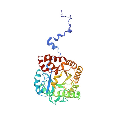
The X-Ray Structure of Yeast 5-Aminolaevulinic Acid Dehydratase Complexed with Substrate and Three Inhibitors

Erskine, P.T.Newbold, R.Brindley, A.A.Wood, S.P.Shoolingin-Jordan, P.M.Warren, M.J.Cooper, J.B.

(2001) J Mol Biol 312: 133

  • DOI: https://doi.org/10.1006/jmbi.2001.4947
  • Primary Citation of Related Structures:  
    1H7N, 1H7O, 1H7P, 1H7R

  • PubMed Abstract: 

    The structures of 5-aminolaevulinic acid dehydratase (ALAD) complexed with substrate (5-aminolaevulinic acid) and three inhibitors: laevulinic acid, succinylacetone and 4-keto-5-aminolaevulinic acid, have been solved at high resolution. The ligands all bind by forming a covalent link with Lys263 at the active site. The structures define the interactions made by one of the two substrate moieties that bind to the enzyme during catalysis. All of the inhibitors induce a significant ordering of the flap covering the active site. Succinylacetone appears to be unique by inducing a number of conformational changes in loops covering the active site, which may be important for understanding the co-operative properties of ALAD enzymes. Succinylacetone is produced in large amounts by patients suffering from the hereditary disease type I tyrosinaemia and its potent inhibition of ALAD also has implications for the pathology of this disease. The most intriguing result is that obtained with 4-keto-5-amino-hexanoic acid, which seems to form a stable carbinolamine intermediate with Lys263. It appears that we have defined the structure of an intermediate of Schiff base formation that the substrate forms upon binding to the P-site of the enzyme.


  • Organizational Affiliation

    Division of Biochemistry and Molecular Biology, School of Biological Sciences, University of Southampton, Bassett Crescent East, Southampton SO16 7PX, UK.


Macromolecules
Find similar proteins by:  (by identity cutoff)  |  3D Structure
Entity ID: 1
MoleculeChains Sequence LengthOrganismDetailsImage
5-AMINOLAEVULINIC ACID DEHYDRATASE341Saccharomyces cerevisiaeMutation(s): 0 
EC: 4.2.1.24
UniProt
Find proteins for P05373 (Saccharomyces cerevisiae (strain ATCC 204508 / S288c))
Explore P05373 
Go to UniProtKB:  P05373
Entity Groups  
Sequence Clusters30% Identity50% Identity70% Identity90% Identity95% Identity100% Identity
UniProt GroupP05373
Sequence Annotations
Expand
  • Reference Sequence
Small Molecules
Experimental Data & Validation

Experimental Data

  • Method: X-RAY DIFFRACTION
  • Resolution: 1.75 Å
  • R-Value Free: 0.353 
  • R-Value Observed: 0.259 
  • Space Group: I 4 2 2
Unit Cell:
Length ( Å )Angle ( ˚ )
a = 102.8α = 90
b = 102.8β = 90
c = 168.3γ = 90
Software Package:
Software NamePurpose
SHELXrefinement
MOSFLMdata reduction
Agrovatadata scaling
SCALAdata scaling
CCP4phasing

Structure Validation

View Full Validation Report


Entry History 

Deposition Data

Revision History  (Full details and data files)

  • Version 1.0: 2001-07-10
    Type: Initial release
  • Version 1.1: 2011-05-08
    Changes: Version format compliance
  • Version 1.2: 2011-07-13
    Changes: Version format compliance
  • Version 1.3: 2019-05-22
    Changes: Data collection, Derived calculations, Other, Refinement description
  • Version 1.4: 2019-07-24
    Changes: Data collection
  • Version 2.0: 2023-11-15
    Changes: Atomic model, Data collection, Database references, Derived calculations, Other
  • Version 2.1: 2023-12-13
    Changes: Refinement description