Refinement of the Spatial Structure of the Gramicidin a Ion Channel
Lomize, A.L., Orekhov, V.I.U., Arsen'Ev, A.S.(1992) Биологические мембраны Журнал мембранной и клеточной биологии 18: 182
- PubMed: 1376600
- Primary Citation of Related Structures:
1GRM - PubMed Abstract:
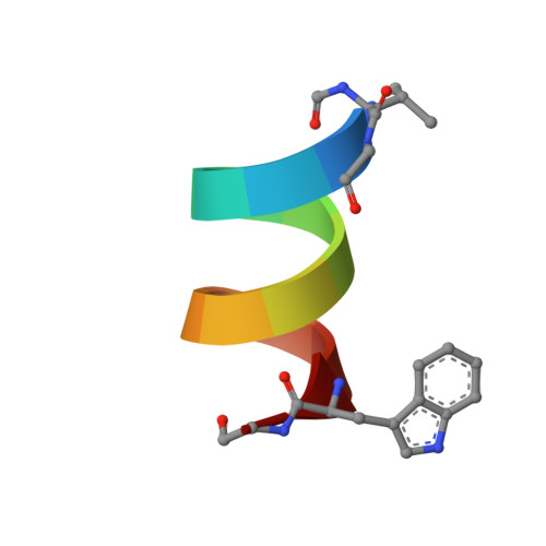
The spatial structure of the gramicidin A (GA) transmembrane ion-channel was refined on the base of cross-peak volumes measured in NOESY spectra (mixing time tau m = 100 and 200 ms). The refinement methods included the comparison of experimental cross-peak volumes with those calculated for low-energy GA conformations, dynamic averaging of the low-energy conformation set and restrained energy minimization. Accuracy of the spatial structure determination was estimated by the penalty function Fr defined as a root mean square deviation of interproton distances corresponding to the calculated and experimental cross-peak volumes. As the initial conformation we used the right-handed pi 6,3 LD pi 6,3 LD helix established on the base of NMR data regardless of the cross-peak volumes. The conformation is in a good agreement with NOE cross-peak volumes (Fr 0.2 to 0.5 A depending on NOESY spectrum). For a number of NOEs formed by the side chain protons, distances errors were found as much as 0.5-2.0 A. Restrained energy minimization procedure had little further success. However some of these errors were eliminated by the change in torsional angle chi 2 of D-Leu12 and dynamic averaging of the Val7 side chain conformations. Apparently, majority of deviations of the calculated and experimental cross-peak volumes are due to the intramolecular mobility of GA and cannot be eliminated within the framework of rigid globule model. In summary the spatial structure of GA ion-channel can be thought as a set of low-energy conformations, differing by the side chain torsion angles chi 1 Val7 and chi 2 D-Leu4 and D-Leu10 and the orientation of the C-terminal ethanolamine group. Root mean square differences between the atomic coordinates of conformations are in the range of 0.3-0.8 A.