1GFO

OMPF PORIN (MUTANT R132P)


Experimental Data Snapshot

  • Method: X-RAY DIFFRACTION
  • Resolution: 3.30 Å
  • R-Value Work: 0.190 
  • R-Value Observed: 0.190 

wwPDB Validation   3D Report Full Report


Ligand Structure Quality Assessment 


This is version 1.5 of the entry. See complete history


Literature

Structural and functional characterization of OmpF porin mutants selected for larger pore size. I. Crystallographic analysis.

Lou, K.L.Saint, N.Prilipov, A.Rummel, G.Benson, S.A.Rosenbusch, J.P.Schirmer, T.

(1996) J Biol Chem 271: 20669-20675

  • Primary Citation of Related Structures:  
    1GFM, 1GFN, 1GFO, 1GFP, 1GFQ

  • PubMed Abstract: 

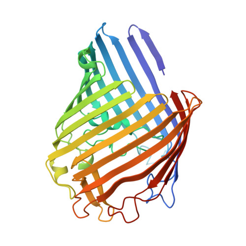
    OmpF porin is a nonspecific pore protein from the outer membrane of Escherichia coli. Previously, a set of mutants was selected that allow the passage of long maltodextrins that do not translocate through the wild-type pore. Here, we describe the crystal structures of four point mutants and one deletion mutant from this set; their functional characterization is reported in the accompanying paper (Saint, N., Lou, K.-L., Widmer, C., Luckey, M., Schirmer, T., Rosenbusch, J. P. (1996) J. Biol. Chem. 271, 20676-20680). All mutations have a local effect on the structure of the pore constriction and result in a larger pore cross-section. Substitution of each of the three closely packed arginine residues at the pore constriction (Arg-42, Arg-82, and Arg-132) by shorter uncharged residues causes rearrangement of the adjacent basic residues. This demonstrates mutual stabilization of these residues in the wild-type porin. Deletion of six residues from the internal loop (Delta109-114) results in disorder of seven adjacent residues but does not alter the structure of the beta-barrel framework. Thus, the large hollow beta-barrel motif can be regarded as an autonomous structure.


  • Organizational Affiliation

    Department of Structural Biology, Biozentrum, University of Basel, Klingelbergstrasse 70, CH-4056 Basel, Switzerland.


Macromolecules
Find similar proteins by:  (by identity cutoff)  |  3D Structure
Entity ID: 1
MoleculeChains Sequence LengthOrganismDetailsImage
MATRIX PORIN OUTER MEMBRANE PROTEIN F340Escherichia coliMutation(s): 1 
Membrane Entity: Yes 
UniProt
Find proteins for P02931 (Escherichia coli (strain K12))
Explore P02931 
Go to UniProtKB:  P02931
Entity Groups  
Sequence Clusters30% Identity50% Identity70% Identity90% Identity95% Identity100% Identity
UniProt GroupP02931
Sequence Annotations
Expand
  • Reference Sequence
Small Molecules
Ligands 1 Unique
IDChains Name / Formula / InChI Key2D Diagram3D Interactions
C8E
Query on C8E

Download Ideal Coordinates CCD File 
B [auth A]
C [auth A]
D [auth A]
E [auth A]
F [auth A]
B [auth A],
C [auth A],
D [auth A],
E [auth A],
F [auth A],
G [auth A],
H [auth A],
I [auth A],
J [auth A],
K [auth A],
L [auth A],
M [auth A]
(HYDROXYETHYLOXY)TRI(ETHYLOXY)OCTANE
C16 H34 O5
FEOZZFHAVXYAMB-UHFFFAOYSA-N
Experimental Data & Validation

Experimental Data

  • Method: X-RAY DIFFRACTION
  • Resolution: 3.30 Å
  • R-Value Work: 0.190 
  • R-Value Observed: 0.190 
  • Space Group: P 3 2 1
Unit Cell:
Length ( Å )Angle ( ˚ )
a = 118.2α = 90
b = 118.2β = 90
c = 52.8γ = 120
Software Package:
Software NamePurpose
X-PLORmodel building
X-PLORrefinement
MADNESdata reduction
X-PLORphasing

Structure Validation

View Full Validation Report


Ligand Structure Quality Assessment 


Entry History 

Deposition Data

Revision History  (Full details and data files)

  • Version 1.0: 1996-12-07
    Type: Initial release
  • Version 1.1: 2008-03-24
    Changes: Version format compliance
  • Version 1.2: 2011-07-13
    Changes: Derived calculations, Version format compliance
  • Version 1.3: 2018-04-18
    Changes: Advisory, Data collection, Other
  • Version 1.4: 2021-11-03
    Changes: Advisory, Database references, Derived calculations
  • Version 1.5: 2024-02-07
    Changes: Data collection