Anatomy of an engineered NAD-binding site.
Mittl, P.R., Berry, A., Scrutton, N.S., Perham, R.N., Schulz, G.E.(1994) Protein Sci 3: 1504-1514
- PubMed: 7833810
- DOI: https://doi.org/10.1002/pro.5560030916
- Primary Citation of Related Structures:
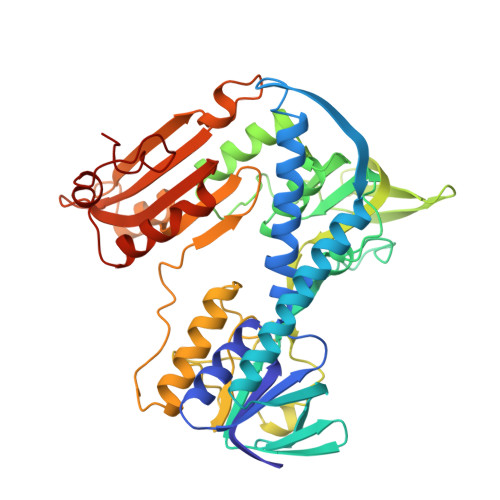
1GES, 1GET, 1GEU - PubMed Abstract:
The coenzyme specificity of Escherichia coli glutathione reductase was switched from NADP to NAD by modifying the environment of the 2'-phosphate binding site through a set of point mutations: A179G, A183G, V197E, R198M, K199F, H200D, and R204P (Scrutton NS, Berry A, Perham RN, 1990, Nature 343:38-43). In order to analyze the structural changes involved, we have determined 4 high-resolution crystal structures, i.e., the structures of the wild-type enzyme (1.86 A resolution, R-factor of 16.8%), of the wild-type enzyme ligated with NADP (2.0 A, 20.8%), of the NAD-dependent mutant (1.74 A, 16.8%), and of the NAD-dependent mutant ligated with NAD (2.2 A, 16.9%). A comparison of these structures reveals subtle differences that explain details of the specificity change. In particular, a peptide rotation occurs close to the adenosine ribose, with a concomitant change of the ribose pucker. The mutations cause a contraction of the local chain fold. Furthermore, the engineered NAD-binding site assumes a less rigid structure than the NADP site of the wild-type enzyme. A superposition of the ligated structures shows a displacement of NAD versus NADP such that the electron pathway from the nicotinamide ring to FAD is elongated, which may explain the lower catalytic efficiency of the mutant. Because the nicotinamide is as much as 15 A from the sites of the mutations, this observation reminds us that mutations may have important long-range consequences that are difficult to anticipate.
Organizational Affiliation:
Institut für Organische Chemie und Biochemie, Albert-Ludwigs-Universität, Freiburg, Germany.