1GBN

HUMAN ORNITHINE AMINOTRANSFERASE COMPLEXED WITH THE NEUROTOXIN GABACULINE


Experimental Data Snapshot

  • Method: X-RAY DIFFRACTION
  • Resolution: 2.30 Å
  • R-Value Free: 0.235 
  • R-Value Work: 0.209 
  • R-Value Observed: 0.209 

wwPDB Validation   3D Report Full Report


Ligand Structure Quality Assessment 


This is version 1.3 of the entry. See complete history


Literature

Human ornithine aminotransferase complexed with L-canaline and gabaculine: structural basis for substrate recognition.

Shah, S.A.Shen, B.W.Brunger, A.T.

(1997) Structure 5: 1067-1075

  • DOI: https://doi.org/10.1016/s0969-2126(97)00258-x
  • Primary Citation of Related Structures:  
    1GBN, 2CAN

  • PubMed Abstract: 

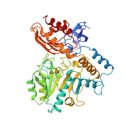
    Ornithine aminotransferase (OAT) is a 45 kDa pyridoxal-5'-phosphate (PLP)-dependent enzyme that catalyzes the conversion of L-ornithine and 2-oxoglutarate to glutamate-delta-semialdehyde and glutamic acid, respectively. In humans, loss of OAT function causes an accumulation of ornithine that results in gyrate atrophy of the choroid and retina, a disease that progressively leads to blindness. In an effort to learn more about the structural basis of this enzyme's function, we have determined the X-ray structures of OAT in complex with two enzyme-activated suicide substrates: L-canaline, an ornithine analog, and gabaculine, an irreversible inhibitor of several related aminotransferases.


  • Organizational Affiliation

    Department of Molecular Biophysics and Biochemistry, Yale University, New Haven, CT 06520, USA.


Macromolecules
Find similar proteins by:  (by identity cutoff)  |  3D Structure
Entity ID: 1
MoleculeChains Sequence LengthOrganismDetailsImage
ORNITHINE AMINOTRANSFERASE
A, B, C
402Homo sapiensMutation(s): 0 
EC: 2.6.1.13
UniProt & NIH Common Fund Data Resources
Find proteins for P04181 (Homo sapiens)
Explore P04181 
Go to UniProtKB:  P04181
PHAROS:  P04181
GTEx:  ENSG00000065154 
Entity Groups  
Sequence Clusters30% Identity50% Identity70% Identity90% Identity95% Identity100% Identity
UniProt GroupP04181
Sequence Annotations
Expand
  • Reference Sequence
Experimental Data & Validation

Experimental Data

  • Method: X-RAY DIFFRACTION
  • Resolution: 2.30 Å
  • R-Value Free: 0.235 
  • R-Value Work: 0.209 
  • R-Value Observed: 0.209 
  • Space Group: P 32 2 1
Unit Cell:
Length ( Å )Angle ( ˚ )
a = 115.013α = 90
b = 115.013β = 90
c = 185.753γ = 120
Software Package:
Software NamePurpose
X-PLORmodel building
X-PLORrefinement
DENZOdata reduction
SCALEPACKdata scaling
X-PLORphasing

Structure Validation

View Full Validation Report


Ligand Structure Quality Assessment 


Entry History 

Deposition Data

Revision History  (Full details and data files)

  • Version 1.0: 1998-06-03
    Type: Initial release
  • Version 1.1: 2008-03-03
    Changes: Version format compliance
  • Version 1.2: 2011-07-13
    Changes: Derived calculations, Version format compliance
  • Version 1.3: 2023-08-09
    Changes: Database references, Derived calculations, Other, Refinement description