1G74

Toward changing specificity: adipocyte lipid binding protein mutant, oleic acid bound form


Experimental Data Snapshot

  • Method: X-RAY DIFFRACTION
  • Resolution: 1.70 Å
  • R-Value Free: 0.238 
  • R-Value Work: 0.208 

wwPDB Validation   3D Report Full Report


Ligand Structure Quality Assessment 


This is version 1.5 of the entry. See complete history


Literature

Specificity determinants for lipids bound to beta-barrel proteins.

Reese, A.J.Banaszak, L.J.

(2004) J Lipid Res 45: 232-243

  • DOI: https://doi.org/10.1194/jlr.M300113-JLR200
  • Primary Citation of Related Structures:  
    1G74, 1G7N

  • PubMed Abstract: 

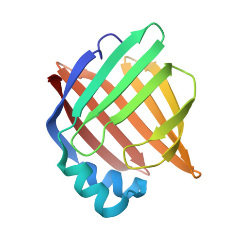
    The family of proteins accountable for the intracellular movement of lipids is characterized by a 10-stranded beta-barrel that forms an internalized cavity varying in size and binding preferences. The loop connecting beta-strands E and F (the fifth and sixth strands) is the most striking conformational difference between adipocyte lipid binding protein (ALBP; fatty acids) and cellular retinoic acid binding protein type I (CRABP I). A three-residue mutation was made in wild-type (WT)-ALBP [ALBP with a three-residue mutation (EF-ALBP)] to mimic CRABP I. Crystal structures of ligand-free and EF-ALBP with bound oleic acid were solved to resolutions of 1.5 A and 1.7 A, respectively, and compared with previous studies of WT-ALBP. The changes in three residues of one loop of the protein appear to have altered the positioning of the C18 fatty acid, as observed in the electron density of EF-ALBP. The crystallographic studies made it possible to compare the protein conformation and ligand positioning with those found in the WT protein. Although the cavity binding sites in both the retinoid and fatty acid binding proteins are irregular, the ligand atoms appear to favor a relatively planar region of the cavities. Preliminary chemical characterization of the mutant protein indicated changes in some binding properties and overall protein stability.


  • Organizational Affiliation

    Department of Molecular Microbiology, Washington University School of Medicine, St. Louis, MO, USA.


Macromolecules
Find similar proteins by:  (by identity cutoff)  |  3D Structure
Entity ID: 1
MoleculeChains Sequence LengthOrganismDetailsImage
ADIPOCYTE LIPID-BINDING PROTEIN131Mus musculusMutation(s): 3 
Gene Names: ALBP
UniProt
Find proteins for P04117 (Mus musculus)
Explore P04117 
Go to UniProtKB:  P04117
Entity Groups  
Sequence Clusters30% Identity50% Identity70% Identity90% Identity95% Identity100% Identity
UniProt GroupP04117
Sequence Annotations
Expand
  • Reference Sequence
Small Molecules
Binding Affinity Annotations 
IDSourceBinding Affinity
OLA BindingDB:  1G74 Ki: min: 185, max: 1500 (nM) from 2 assay(s)
IC50: 2.61e+4 (nM) from 1 assay(s)
Binding MOAD:  1G74 Kd: 300 (nM) from 1 assay(s)
PDBBind:  1G74 Kd: 300 (nM) from 1 assay(s)
Experimental Data & Validation

Experimental Data

  • Method: X-RAY DIFFRACTION
  • Resolution: 1.70 Å
  • R-Value Free: 0.238 
  • R-Value Work: 0.208 
  • Space Group: C 1 2 1
Unit Cell:
Length ( Å )Angle ( ˚ )
a = 118.92α = 90
b = 36.89β = 92.61
c = 27.87γ = 90
Software Package:
Software NamePurpose
CNSrefinement
CNSphasing

Structure Validation

View Full Validation Report


Ligand Structure Quality Assessment 


Entry History 

Deposition Data

Revision History  (Full details and data files)

  • Version 1.0: 2003-06-10
    Type: Initial release
  • Version 1.1: 2008-04-27
    Changes: Version format compliance
  • Version 1.2: 2011-07-13
    Changes: Version format compliance
  • Version 1.3: 2017-10-04
    Changes: Refinement description
  • Version 1.4: 2021-10-27
    Changes: Database references, Derived calculations
  • Version 1.5: 2023-08-09
    Changes: Data collection, Refinement description