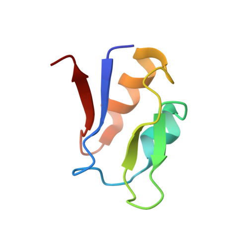
Crystal structure of the ferredoxin I from Desulfovibrio africanus at 2.3 A resolution.
Sery, A., Housset, D., Serre, L., Bonicel, J., Hatchikian, C., Frey, M., Roth, M.(1994) Biochemistry 33: 15408-15417
- PubMed: 7803404
- DOI: https://doi.org/10.1021/bi00255a022
- Primary Citation of Related Structures:
1FXR - PubMed Abstract:
The crystal structure of the ferredoxin I from the sulfate-reducing bacterium Desulfovibrio africanus (DaFdI) has been solved and refined by X-ray diffraction. The crystals are orthorhombic with a = 96.6 A, b = 58.1 A, and c = 20.7 A, space group P2(1)2(1)2, and two ferredoxin molecules per asymmetric unit. The initial electron density map has been obtained by combining phasing by molecular replacement methods, anomalous scattering, and noncrystallographic averaging. The final crystallographic R factor is 0.182 with 10-2.3 A resolution data. In parallel, the amino acid sequence was redetermined. This showed that DaFdI contains 64 residues (instead of 61) including one free cysteine, one histidine, and one tryptophan in the C-terminal part of the molecule. The current molecular model includes the two molecules of the asymmetric unit, 67 water molecules, and one sulfate ion. The DaFdI overall folding very closely resembles that of ferredoxins of known structure. Comparisons with the single cluster ferredoxins from Desulfovibrio gigas and Bacillus thermoproteolyticus show that the presence or the absence of a disulfide bridge does not significantly affect the folding of the other half of the molecule, including the characteristic alpha-helix of the single cluster ferreddoxins. Like other ferredoxins or analogs, the [4Fe-4S] iron--sulfur cluster presents, at 2.3 A resolution, a cubane-like geometry. By contrast, its immediate environment is different as it includes, besides the four cysteic sulfur ligands, the sulfur atom of the free cysteine. This sulfur atom, which is buried within the protein, is in van der Waals contact with one labile sulfur of the cluster and one liganded cysteic sulfur. The association of a [4Fe-4S] cluster with one free cysteic sulfur is similar to that previously found in both X-ray structures of Azotobacter vinelandii and Peptococcus aerogenes [Stout, C. D. (1989) J. Mol. Biol. 205, 545-555; Backes, G., et al. (1991) J. Am. Chem. Soc. 113, 2055-2064]. Chemical sequence analysis suggests that this characteristic [4Fe-4S] cluster sulfur environment is widely distributed among ferredoxins.
Organizational Affiliation:
Laboratoire de Cristallographie et de Cristallogénèse des Protéines, Institut de Biologie Structurale J.-P. Ebel, CEA-CNRS, Grenoble, France.