1FPR

CRYSTAL STRUCTURE OF THE COMPLEX FORMED BETWEEN THE CATALYTIC DOMAIN OF SHP-1 AND AN IN VITRO PEPTIDE SUBSTRATE PY469 DERIVED FROM SHPS-1.


Experimental Data Snapshot

  • Method: X-RAY DIFFRACTION
  • Resolution: 2.50 Å
  • R-Value Free: 0.303 
  • R-Value Work: 0.194 
  • R-Value Observed: 0.199 

wwPDB Validation   3D Report Full Report


This is version 1.4 of the entry. See complete history


Literature

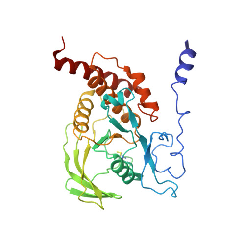
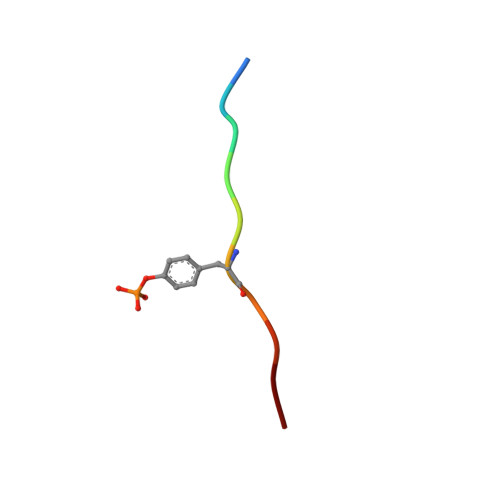
Structural basis for substrate specificity of protein-tyrosine phosphatase SHP-1.

Yang, J.Cheng, Z.Niu, T.Liang, X.Zhao, Z.J.Zhou, G.W.

(2000) J Biol Chem 275: 4066-4071

  • DOI: https://doi.org/10.1074/jbc.275.6.4066
  • Primary Citation of Related Structures:  
    1FPR

  • PubMed Abstract: 

    The substrate specificity of the catalytic domain of SHP-1, an important regulator in the proliferation and development of hematopoietic cells, is critical for understanding the physiological functions of SHP-1. Here we report the crystal structures of the catalytic domain of SHP-1 complexed with two peptide substrates derived from SIRPalpha, a member of the signal-regulatory proteins. We show that the variable beta5-loop-beta6 motif confers SHP-1 substrate specificity at the P-4 and further N-terminal subpockets. We also observe a novel residue shift at P-2, the highly conserved subpocket in protein- tyrosine phosphatases. Our observations provide new insight into the substrate specificity of SHP-1.


  • Organizational Affiliation

    Program in Molecular Medicine, University of Massachusetts Medical School, Worcester, Massachusetts 01605, USA.


Macromolecules
Find similar proteins by:  (by identity cutoff)  |  3D Structure
Entity ID: 1
MoleculeChains Sequence LengthOrganismDetailsImage
PROTEIN-TYROSINE PHOSPHATASE 1C284Homo sapiensMutation(s): 1 
EC: 3.1.3.48
UniProt & NIH Common Fund Data Resources
Find proteins for P29350 (Homo sapiens)
Explore P29350 
Go to UniProtKB:  P29350
PHAROS:  P29350
GTEx:  ENSG00000111679 
Entity Groups  
Sequence Clusters30% Identity50% Identity70% Identity90% Identity95% Identity100% Identity
UniProt GroupP29350
Sequence Annotations
Expand
  • Reference Sequence

Find similar proteins by:  Sequence   |   3D Structure  

Entity ID: 2
MoleculeChains Sequence LengthOrganismDetailsImage
PEPTIDE PY46910N/AMutation(s): 0 
Entity Groups  
Sequence Clusters30% Identity50% Identity70% Identity90% Identity95% Identity100% Identity
Sequence Annotations
Expand
  • Reference Sequence
Small Molecules
Modified Residues  1 Unique
IDChains TypeFormula2D DiagramParent
PTR
Query on PTR
B
L-PEPTIDE LINKINGC9 H12 N O6 PTYR
Experimental Data & Validation

Experimental Data

  • Method: X-RAY DIFFRACTION
  • Resolution: 2.50 Å
  • R-Value Free: 0.303 
  • R-Value Work: 0.194 
  • R-Value Observed: 0.199 
  • Space Group: P 21 21 2
Unit Cell:
Length ( Å )Angle ( ˚ )
a = 111.58α = 90
b = 45.21β = 90
c = 56.27γ = 90
Software Package:
Software NamePurpose
MAR345data collection
SCALEPACKdata scaling
AMoREphasing
X-PLORrefinement

Structure Validation

View Full Validation Report


Entry History 

Deposition Data

Revision History  (Full details and data files)

  • Version 1.0: 2001-03-07
    Type: Initial release
  • Version 1.1: 2008-04-27
    Changes: Version format compliance
  • Version 1.2: 2011-07-13
    Changes: Version format compliance
  • Version 1.3: 2017-10-04
    Changes: Refinement description
  • Version 1.4: 2021-11-03
    Changes: Database references, Derived calculations