1FNH

CRYSTAL STRUCTURE OF HEPARIN AND INTEGRIN BINDING SEGMENT OF HUMAN FIBRONECTIN


Experimental Data Snapshot

  • Method: X-RAY DIFFRACTION
  • Resolution: 2.80 Å
  • R-Value Free: 0.295 
  • R-Value Work: 0.216 

wwPDB Validation   3D Report Full Report


This is version 1.3 of the entry. See complete history


Literature

Crystal structure of a heparin- and integrin-binding segment of human fibronectin.

Sharma, A.Askari, J.A.Humphries, M.J.Jones, E.Y.Stuart, D.I.

(1999) EMBO J 18: 1468-1479

  • DOI: https://doi.org/10.1093/emboj/18.6.1468
  • Primary Citation of Related Structures:  
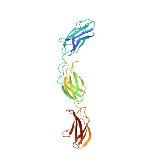
    1FNH

  • PubMed Abstract: 

    The crystal structure of human fibronectin (FN) type III repeats 12-14 reveals the primary heparin-binding site, a clump of positively charged residues in FN13, and a putative minor site approximately 60 A away in FN14. The IDAPS motif implicated in integrin alpha4beta1 binding is at the FN13-14 junction, rendering the critical Asp184 inaccessible to integrin. Asp184 clamps the BC loop of FN14, whose sequence (PRARI) is reminiscent of the synergy sequence (PHSRN) of FN9. Mutagenesis studies prompted by this observation reveal that both arginines of the PRARI sequence are important for alpha4beta1 binding to FN12-14. The PRARI motif may represent a new class of integrin-binding sites. The spatial organization of the binding sites suggests that heparin and integrin may bind in concert.


  • Organizational Affiliation

    Laboratory of Molecular Biophysics, University of Oxford, Rex Richardson Building, South Parks Road, Oxford, OX1 3QU, UK.


Macromolecules
Find similar proteins by:  (by identity cutoff)  |  3D Structure
Entity ID: 1
MoleculeChains Sequence LengthOrganismDetailsImage
PROTEIN (FIBRONECTIN)271Homo sapiensMutation(s): 0 
UniProt & NIH Common Fund Data Resources
Find proteins for P02751 (Homo sapiens)
Explore P02751 
Go to UniProtKB:  P02751
PHAROS:  P02751
GTEx:  ENSG00000115414 
Entity Groups  
Sequence Clusters30% Identity50% Identity70% Identity90% Identity95% Identity100% Identity
UniProt GroupP02751
Sequence Annotations
Expand
  • Reference Sequence
Experimental Data & Validation

Experimental Data

  • Method: X-RAY DIFFRACTION
  • Resolution: 2.80 Å
  • R-Value Free: 0.295 
  • R-Value Work: 0.216 
  • Space Group: I 2 2 2
Unit Cell:
Length ( Å )Angle ( ˚ )
a = 68.58α = 90
b = 86.29β = 90
c = 142.8γ = 90
Software Package:
Software NamePurpose
CCP4model building
SHARPphasing
CNSrefinement
DENZOdata reduction
SCALEPACKdata scaling
CCP4phasing

Structure Validation

View Full Validation Report


Entry History 

Deposition Data

Revision History  (Full details and data files)

  • Version 1.0: 1999-03-16
    Type: Initial release
  • Version 1.1: 2008-04-26
    Changes: Version format compliance
  • Version 1.2: 2011-07-13
    Changes: Version format compliance
  • Version 1.3: 2023-12-27
    Changes: Data collection, Database references