1EB0

Crystal structure of Bacillus pasteurii UreE at 1.85 A, phased by SIRAS. Type I crystal form.


Experimental Data Snapshot

  • Method: X-RAY DIFFRACTION
  • Resolution: 1.85 Å
  • R-Value Free: 0.256 
  • R-Value Work: 0.220 
  • R-Value Observed: 0.220 

wwPDB Validation   3D Report Full Report


This is version 1.6 of the entry. See complete history


Literature

Structural Basis for Ni2+ Transport and Assembly of the Urease Active Site by the Metallochaperone Uree from Bacillus Pasteurii

Reamut, H.Safarov, N.Ciurli, S.Van Beeumen, J.

(2001) J Biol Chem 276: 49365

  • DOI: https://doi.org/10.1074/jbc.M108304200
  • Primary Citation of Related Structures:  
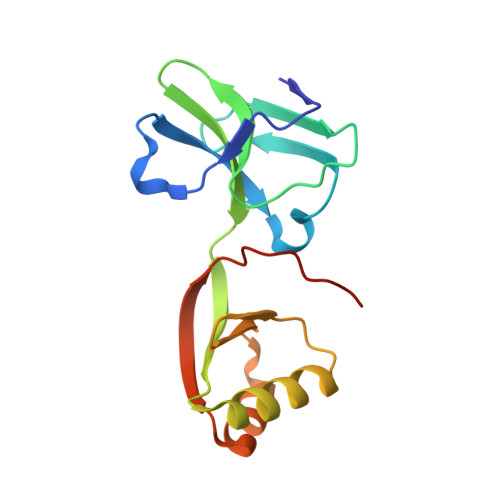
    1EAR, 1EB0

  • PubMed Abstract: 

    Bacillus pasteurii UreE (BpUreE) is a putative chaperone assisting the insertion of Ni(2+) ions in the active site of urease. The x-ray structure of the protein has been determined for two crystal forms, at 1.7 and 1.85 A resolution, using SIRAS phases derived from a Hg(2+)-derivative. BpUreE is composed of distinct N- and C-terminal domains, connected by a short flexible linker. The structure reveals the topology of an elongated homodimer, formed by interaction of the two C-terminal domains through hydrophobic interactions. A single Zn(2+) ion bound to four conserved His-100 residues, one from each monomer, connects two dimers resulting in a tetrameric BpUreE known to be formed in concentrated solutions. The Zn(2+) ion can be replaced by Ni(2+) as shown by anomalous difference maps obtained on a crystal of BpUreE soaked in a solution containing NiCl(2). A large hydrophobic patch surrounding the metal ion site is surface-exposed in the biologically relevant dimer. The BpUreE structure represents the first for this class of proteins and suggests a possible role for UreE in the urease nickel-center assembly.


  • Organizational Affiliation

    Laboratory of Protein Biochemistry and Protein Engineering, Ghent University, K. L. Ledeganckstraat 35, B-9000 Gent, Belgium.


Macromolecules
Find similar proteins by:  (by identity cutoff)  |  3D Structure
Entity ID: 1
MoleculeChains Sequence LengthOrganismDetailsImage
UREASE ACCESSORY PROTEIN UREE147Sporosarcina pasteuriiMutation(s): 0 
UniProt
Find proteins for P50049 (Sporosarcina pasteurii)
Explore P50049 
Go to UniProtKB:  P50049
Entity Groups  
Sequence Clusters30% Identity50% Identity70% Identity90% Identity95% Identity100% Identity
UniProt GroupP50049
Sequence Annotations
Expand
  • Reference Sequence
Small Molecules
Ligands 1 Unique
IDChains Name / Formula / InChI Key2D Diagram3D Interactions
ZN
Query on ZN

Download Ideal Coordinates CCD File 
B [auth A]ZINC ION
Zn
PTFCDOFLOPIGGS-UHFFFAOYSA-N
Experimental Data & Validation

Experimental Data

  • Method: X-RAY DIFFRACTION
  • Resolution: 1.85 Å
  • R-Value Free: 0.256 
  • R-Value Work: 0.220 
  • R-Value Observed: 0.220 
  • Space Group: I 2 2 2
Unit Cell:
Length ( Å )Angle ( ˚ )
a = 50.5α = 90
b = 60.72β = 90
c = 130.58γ = 90
Software Package:
Software NamePurpose
CNSrefinement
DENZOdata reduction
SCALEPACKdata scaling
MLPHAREphasing

Structure Validation

View Full Validation Report


Entry History 

Deposition Data

Revision History  (Full details and data files)

  • Version 1.0: 2002-01-04
    Type: Initial release
  • Version 1.1: 2011-05-08
    Changes: Version format compliance
  • Version 1.2: 2011-07-13
    Changes: Version format compliance
  • Version 1.3: 2018-06-27
    Changes: Data collection, Derived calculations
  • Version 1.4: 2019-05-08
    Changes: Data collection, Experimental preparation
  • Version 1.5: 2019-07-10
    Changes: Data collection
  • Version 1.6: 2019-07-24
    Changes: Data collection