Structural Analysis of Interactions for Complex Formation between Ferredoxin-Nadp+ Reductase and its Protein Partners.
Mayoral, T., Martinez-Julvez, M., Perez-Dorado, I., Sanz-Aparicio, J., Gomez-Moreno, C., Medina, M., Hermoso, J.A.(2005) Proteins 59: 592
- PubMed: 15789405
- DOI: https://doi.org/10.1002/prot.20450
- Primary Citation of Related Structures:
1E62, 1E63, 1E64, 1GO2, 1QGY - PubMed Abstract:
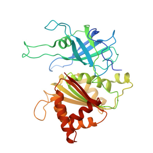
The three-dimensional structures of K72E, K75R, K75S, K75Q, and K75E Anabaena Ferredoxin-NADP+ reductase (FNR) mutants have been solved, and particular structural details of these mutants have been used to assess the role played by residues 72 and 75 in optimal complex formation and electron transfer (ET) between FNR and its protein redox partners Ferredoxin (Fd) and Flavodoxin (Fld). Additionally, because there is no structural information available on the interaction between FNR and Fld, a model for the FNR:Fld complex has also been produced based on the previously reported crystal structures and on that of the rat Cytochrome P450 reductase (CPR), onto which FNR and Fld have been structurally aligned, and those reported for the Anabaena and maize FNR:Fd complexes. The model suggests putative electrostatic and hydrophobic interactions between residues on the FNR and Fld surfaces at the complex interface and provides an adequate orientation and distance between the FAD and FMN redox centers for efficient ET without the presence of any other molecule as electron carrier. Thus, the models now available for the FNR:Fd and FNR:Fld interactions and the structures presented here for the mutants at K72 and K75 in Anabaena FNR have been evaluated in light of previous biochemical data. These structures confirm the key participation of residue K75 and K72 in complex formation with both Fd and Fld. The drastic effect in FNR activity produced by replacement of K75 by Glu in the K75E FNR variant is explained not only by the observed changes in the charge distribution on the surface of the K75E FNR mutant, but also by the formation of a salt bridge interaction between E75 and K72 that simultaneously "neutralizes" two essential positive charged side chains for Fld/Fd recognition.
Organizational Affiliation:
Grupo de Cristalografía Macromolecular y Biología Estructural, Instituto Química-Física Rocasolano, C.S.I.C. Serrano 119, 28006-Madrid, Spain.