1E3G

Human Androgen Receptor Ligand Binding in complex with the ligand metribolone (R1881)


Experimental Data Snapshot

  • Method: X-RAY DIFFRACTION
  • Resolution: 2.40 Å
  • R-Value Free: 0.297 
  • R-Value Work: 0.210 
  • R-Value Observed: 0.210 

wwPDB Validation   3D Report Full Report


Ligand Structure Quality Assessment 


This is version 2.2 of the entry. See complete history


Literature

Structural evidence for ligand specificity in the binding domain of the human androgen receptor. Implications for pathogenic gene mutations.

Matias, P.M.Donner, P.Coelho, R.Thomaz, M.Peixoto, C.Macedo, S.Otto, N.Joschko, S.Scholz, P.Wegg, A.Basler, S.Schafer, M.Egner, U.Carrondo, M.A.

(2000) J Biol Chem 275: 26164-26171

  • DOI: https://doi.org/10.1074/jbc.M004571200
  • Primary Citation of Related Structures:  
    1E3G, 1E3K

  • PubMed Abstract: 

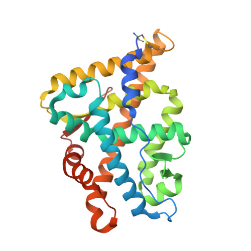
    The crystal structures of the human androgen receptor (hAR) and human progesterone receptor ligand-binding domains in complex with the same ligand metribolone (R1881) have been determined. Both three-dimensional structures show the typical nuclear receptor fold. The change of two residues in the ligand-binding pocket between the human progesterone receptor and hAR is most likely the source for the specificity of R1881 to the hAR. The structural implications of the 14 known mutations in the ligand-binding pocket of the hAR ligand-binding domains associated with either prostate cancer or the partial or complete androgen receptor insensitivity syndrome were analyzed. The effects of most of these mutants could be explained on the basis of the crystal structure.


  • Organizational Affiliation

    Instituto de Tecnologia Quimica e Biológica, Universidade Nova de Lisboa, Apartado 127, 2780 Oeiras, Portugal.


Macromolecules
Find similar proteins by:  (by identity cutoff)  |  3D Structure
Entity ID: 1
MoleculeChains Sequence LengthOrganismDetailsImage
ANDROGEN RECEPTOR263Homo sapiensMutation(s): 0 
UniProt & NIH Common Fund Data Resources
Find proteins for P10275 (Homo sapiens)
Explore P10275 
Go to UniProtKB:  P10275
PHAROS:  P10275
GTEx:  ENSG00000169083 
Entity Groups  
Sequence Clusters30% Identity50% Identity70% Identity90% Identity95% Identity100% Identity
UniProt GroupP10275
Sequence Annotations
Expand
  • Reference Sequence
Small Molecules
Ligands 1 Unique
IDChains Name / Formula / InChI Key2D Diagram3D Interactions
R18
Query on R18

Download Ideal Coordinates CCD File 
B [auth A](17BETA)-17-HYDROXY-17-METHYLESTRA-4,9,11-TRIEN-3-ONE
C19 H24 O2
CCCIJQPRIXGQOE-XWSJACJDSA-N
Binding Affinity Annotations 
IDSourceBinding Affinity
R18 BindingDB:  1E3G Ki: 0.4 (nM) from 1 assay(s)
Kd: min: 0.52, max: 1.95 (nM) from 10 assay(s)
IC50: min: 0.3, max: 7 (nM) from 5 assay(s)
EC50: 0.01 (nM) from 1 assay(s)
PDBBind:  1E3G Kd: 0.99 (nM) from 1 assay(s)
Experimental Data & Validation

Experimental Data

  • Method: X-RAY DIFFRACTION
  • Resolution: 2.40 Å
  • R-Value Free: 0.297 
  • R-Value Work: 0.210 
  • R-Value Observed: 0.210 
  • Space Group: P 21 21 21
Unit Cell:
Length ( Å )Angle ( ˚ )
a = 54.28α = 90
b = 66.14β = 90
c = 71.72γ = 90
Software Package:
Software NamePurpose
X-PLORrefinement
DENZOdata reduction
SCALEPACKdata scaling
AMoREphasing

Structure Validation

View Full Validation Report


Ligand Structure Quality Assessment 


Entry History 

Revision History  (Full details and data files)

  • Version 1.0: 2001-06-14
    Type: Initial release
  • Version 1.1: 2011-05-08
    Changes: Version format compliance
  • Version 1.2: 2011-07-13
    Changes: Version format compliance
  • Version 2.0: 2018-02-28
    Changes: Advisory, Atomic model, Database references, Source and taxonomy, Structure summary
  • Version 2.1: 2019-05-08
    Changes: Data collection, Experimental preparation
  • Version 2.2: 2023-12-13
    Changes: Advisory, Data collection, Database references, Other, Refinement description