1DOA

Structure of the rho family gtp-binding protein cdc42 in complex with the multifunctional regulator rhogdi


Experimental Data Snapshot

  • Method: X-RAY DIFFRACTION
  • Resolution: 2.60 Å
  • R-Value Free: 0.320 
  • R-Value Work: 0.257 
  • R-Value Observed: 0.257 

wwPDB Validation   3D Report Full Report


This is version 1.2 of the entry. See complete history


Literature

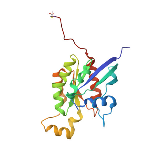
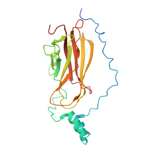
Structure of the Rho family GTP-binding protein Cdc42 in complex with the multifunctional regulator RhoGDI.

Hoffman, G.R.Nassar, N.Cerione, R.A.

(2000) Cell 100: 345-356

  • DOI: https://doi.org/10.1016/s0092-8674(00)80670-4
  • Primary Citation of Related Structures:  
    1DOA

  • PubMed Abstract: 

    The RhoGDI proteins serve as key multifunctional regulators of Rho family GTP-binding proteins. The 2.6 A X-ray crystallographic structure of the Cdc42/RhoGDI complex reveals two important sites of interaction between GDI and Cdc42. First, the amino-terminal regulatory arm of the GDI binds to the switch I and II domains of Cdc42 leading to the inhibition of both GDP dissociation and GTP hydrolysis. Second, the geranylgeranyl moiety of Cdc42 inserts into a hydrophobic pocket within the immunoglobulin-like domain of the GDI molecule leading to membrane release. The structural data demonstrate how GDIs serve as negative regulators of small GTP-binding proteins and how the isoprenoid moiety is utilized in this critical regulatory interaction.


  • Organizational Affiliation

    Department of Molecular Medicine, Veterinary Medical Center, Cornell University, Ithaca, New York 14853, USA.


Macromolecules
Find similar proteins by:  (by identity cutoff)  |  3D Structure
Entity ID: 1
MoleculeChains Sequence LengthOrganismDetailsImage
PROTEIN (GTP-BINDING PROTEIN)191Homo sapiensMutation(s): 1 
Membrane Entity: Yes 
UniProt & NIH Common Fund Data Resources
Find proteins for P60953 (Homo sapiens)
Explore P60953 
Go to UniProtKB:  P60953
PHAROS:  P60953
GTEx:  ENSG00000070831 
Entity Groups  
Sequence Clusters30% Identity50% Identity70% Identity90% Identity95% Identity100% Identity
UniProt GroupP60953
Sequence Annotations
Expand
  • Reference Sequence
Find similar proteins by:  (by identity cutoff)  |  3D Structure
Entity ID: 2
MoleculeChains Sequence LengthOrganismDetailsImage
PROTEIN (GDP-DISSOCIATION INHIBITOR 1)219Bos taurusMutation(s): 0 
UniProt
Find proteins for P19803 (Bos taurus)
Explore P19803 
Go to UniProtKB:  P19803
Entity Groups  
Sequence Clusters30% Identity50% Identity70% Identity90% Identity95% Identity100% Identity
UniProt GroupP19803
Sequence Annotations
Expand
  • Reference Sequence
Small Molecules
Ligands 3 Unique
IDChains Name / Formula / InChI Key2D Diagram3D Interactions
GDP
Query on GDP

Download Ideal Coordinates CCD File 
E [auth A]GUANOSINE-5'-DIPHOSPHATE
C10 H15 N5 O11 P2
QGWNDRXFNXRZMB-UUOKFMHZSA-N
GER
Query on GER

Download Ideal Coordinates CCD File 
F [auth A]GERAN-8-YL GERAN
C20 H34
HSOYJGBJQAKCNA-CAIKYXSQSA-N
MG
Query on MG

Download Ideal Coordinates CCD File 
C [auth A],
D [auth A]
MAGNESIUM ION
Mg
JLVVSXFLKOJNIY-UHFFFAOYSA-N
Modified Residues  1 Unique
IDChains TypeFormula2D DiagramParent
CMT
Query on CMT
A
L-PEPTIDE LINKINGC4 H9 N O2 SCYS
Experimental Data & Validation

Experimental Data

  • Method: X-RAY DIFFRACTION
  • Resolution: 2.60 Å
  • R-Value Free: 0.320 
  • R-Value Work: 0.257 
  • R-Value Observed: 0.257 
  • Space Group: H 3
Unit Cell:
Length ( Å )Angle ( ˚ )
a = 83.94α = 90
b = 83.94β = 90
c = 191.16γ = 120
Software Package:
Software NamePurpose
CNSrefinement
MOSFLMdata reduction
CCP4data scaling
CNSphasing

Structure Validation

View Full Validation Report


Entry History 

Deposition Data

Revision History  (Full details and data files)

  • Version 1.0: 2000-02-09
    Type: Initial release
  • Version 1.1: 2008-04-27
    Changes: Version format compliance
  • Version 1.2: 2011-07-13
    Changes: Version format compliance