Involvement of electrostatic interactions in the mechanism of peptide folding induced by sodium dodecyl sulfate binding.
Montserret, R., McLeish, M.J., Bockmann, A., Geourjon, C., Penin, F.(2000) Biochemistry 39: 8362-8373
- PubMed: 10913242
- DOI: https://doi.org/10.1021/bi000208x
- Primary Citation of Related Structures:
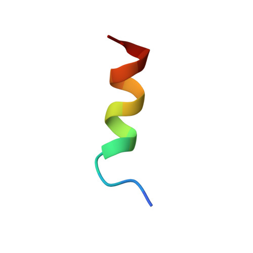
1DJF, 1DN3, 1DNG - PubMed Abstract:
Sodium dodecyl sulfate (SDS) has consistently been shown to induce secondary structure, particularly alpha-helices, in polypeptides, and is commonly used to model membrane and other hydrophobic environments. However, the precise mechanism by which SDS induces these conformational changes remains unclear. To examine the role of electrostatic interactions in this mechanism, we have designed two hydrophilic, charged amphipathic alpha-helical peptides, one basic (QAPAYKKAAKKLAES) and the other acidic (QAPAYEEAAEELAKS), and their structures were studied by CD and NMR. The design of the peptides is based on the sequence of the segment of residues 56-70 of human platelet factor 4 [PF4(56-70), QAPLYKKIIKKLLES]. Both peptides were unstructured in water, and in the presence of neutral, zwitterionic, or cationic detergents. However, in SDS at neutral pH, the basic peptide folded into an alpha-helix. By contrast, the pH needed to be lowered to 1.8 before alpha-helix formation was observed for the acidic peptide. Strong, attractive electrostatic interactions, between the anionic groups of SDS and the cationic groups of the lysines, appeared to be necessary to initiate the folding of the basic peptide. NMR analysis showed that the basic peptide was fully embedded in SDS-peptide micelles, and that its three-dimensional alpha-helical structure could be superimposed on that of the native structure of PF4(56-70). These results enabled us to propose a working model of the basic peptide-SDS complex, and a mechanism for SDS-induced alpha-helical folding. This study demonstrates that, while the folding of peptides is mostly driven by hydrophobic effects, electrostatic interactions play a significant role in the formation and the stabilization of SDS-induced structure.
Organizational Affiliation:
Institut de Biologie et de Chimie des Protéines, CNRS UPR 412, 7 passage du Vercors, Lyon, France.