1DJO

Crystal structure of Pseudomonas 7A Glutaminase-asparaginase with the inhibitor donv covalently bound in the active site


Experimental Data Snapshot

  • Method: X-RAY DIFFRACTION
  • Resolution: 2.00 Å
  • R-Value Free: 0.238 
  • R-Value Work: 0.214 
  • R-Value Observed: 0.214 

wwPDB Validation   3D Report Full Report


This is version 1.2 of the entry. See complete history


Literature

Reactions of Pseudomonas 7A glutaminase-asparaginase with diazo analogues of glutamine and asparagine result in unexpected covalent inhibitions and suggests an unusual catalytic triad Thr-Tyr-Glu.

Ortlund, E.Lacount, M.W.Lewinski, K.Lebioda, L.

(2000) Biochemistry 39: 1199-1204

  • DOI: https://doi.org/10.1021/bi991797d
  • Primary Citation of Related Structures:  
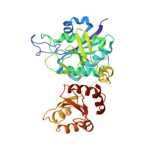
    1DJO, 1DJP

  • PubMed Abstract: 

    Pseudomonas 7A glutaminase-asparaginase (PGA) catalyzes the hydrolysis of D and L isomers of glutamine and asparagine. Crystals of PGA were reacted with diazo analogues of glutamine (6-diazo-5-oxo-L-norleucine, DON) and asparagine (5-diazo-4-oxo-L-norvaline, DONV), which are known inhibitors of the enzyme. The derivatized crystals remained isomorphous to native PGA crystals. Their structures were refined to crystallographic R = 0.20 and R(free) = 0.24 for PGA-DON and R = 0.19 and R = 0.23 for PGA-DONV. Difference Fourier electron density maps clearly showed that both DON and DONV inactivate PGA through covalent inhibition. Continuous electron density connecting the inhibitor to both Thr20 and Tyr34 of the flexible loop was observed providing strong evidence that Thr20 is the primary catalytic nucleophile and that Tyr34 plays an important role in catalysis as well. The unexpected covalent binding observed in the PGA-DON and PGA-DONV complexes shows that a secondary reaction involving the formation of a Tyr34-inhibitor bond takes place with concomitant inactivation of PGA. The predicted covalent linkage is not seen, however, suggesting an alternative method of inhibition not yet seen for these diazo analogues. These surprising results give insight as to the role of the flexible loop Thr and Tyr in the catalytic mechanism.


  • Organizational Affiliation

    Department of Chemistry and Biochemistry, University of South Carolina, Columbia, South Carolina 29208, USA.


Macromolecules
Find similar proteins by:  (by identity cutoff)  |  3D Structure
Entity ID: 1
MoleculeChains Sequence LengthOrganismDetailsImage
GLUTAMINASE-ASPARAGINASE
A, B
330Pseudomonas sp. 7AMutation(s): 0 
EC: 3.5.1.38
UniProt
Find proteins for P10182 (Pseudomonas sp. (strain ATCC 29598 / 7A))
Explore P10182 
Go to UniProtKB:  P10182
Entity Groups  
Sequence Clusters30% Identity50% Identity70% Identity90% Identity95% Identity100% Identity
UniProt GroupP10182
Sequence Annotations
Expand
  • Reference Sequence
Small Molecules
Ligands 1 Unique
IDChains Name / Formula / InChI Key2D Diagram3D Interactions
CAB
Query on CAB

Download Ideal Coordinates CCD File 
C [auth A],
D [auth B]
4,4-dihydroxy-5-oxo-L-norvaline
C5 H9 N O5
NKTBKFGHLWDKLW-VKHMYHEASA-N
Experimental Data & Validation

Experimental Data

  • Method: X-RAY DIFFRACTION
  • Resolution: 2.00 Å
  • R-Value Free: 0.238 
  • R-Value Work: 0.214 
  • R-Value Observed: 0.214 
  • Space Group: C 2 2 21
Unit Cell:
Length ( Å )Angle ( ˚ )
a = 78.37α = 90
b = 135.89β = 90
c = 137.56γ = 90
Software Package:
Software NamePurpose
CNSrefinement
DENZOdata reduction
SCALEPACKdata scaling
CNSphasing

Structure Validation

View Full Validation Report


Entry History 

Deposition Data

Revision History  (Full details and data files)

  • Version 1.0: 2000-01-24
    Type: Initial release
  • Version 1.1: 2008-04-27
    Changes: Version format compliance
  • Version 1.2: 2011-07-13
    Changes: Derived calculations, Source and taxonomy, Version format compliance