1D2S

CRYSTAL STRUCTURE OF THE N-TERMINAL LAMININ G-LIKE DOMAIN OF SHBG IN COMPLEX WITH DIHYDROTESTOSTERONE


Experimental Data Snapshot

  • Method: X-RAY DIFFRACTION
  • Resolution: 1.55 Å
  • R-Value Free: 0.251 
  • R-Value Work: 0.205 
  • R-Value Observed: 0.210 

wwPDB Validation   3D Report Full Report


Ligand Structure Quality Assessment 


This is version 1.4 of the entry. See complete history


Literature

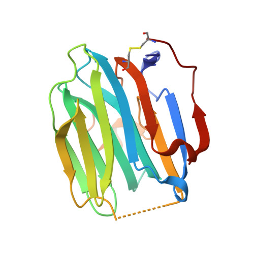
Crystal structure of human sex hormone-binding globulin: steroid transport by a laminin G-like domain.

Grishkovskaya, I.Avvakumov, G.V.Sklenar, G.Dales, D.Hammond, G.L.Muller, Y.A.

(2000) EMBO J 19: 504-512

  • DOI: https://doi.org/10.1093/emboj/19.4.504
  • Primary Citation of Related Structures:  
    1D2S

  • PubMed Abstract: 

    Human sex hormone-binding globulin (SHBG) transports sex steroids in blood and regulates their access to target tissues. In biological fluids, SHBG exists as a homodimer and each monomer comprises two laminin G-like domains (G domains). The crystal structure of the N-terminal G domain of SHBG in complex with 5alpha-dihydrotestosterone at 1.55 A resolution reveals both the architecture of the steroid-binding site and the quaternary structure of the dimer. We also show that G domains have jellyroll topology and are structurally related to pentraxin. In each SHBG monomer, the steroid intercalates into a hydrophobic pocket within the beta-sheet sandwich. The steroid and a 20 A distant calcium ion are not located at the dimer interface. Instead, two separate steroid-binding pockets and calcium-binding sites exist per dimer. The structure displays intriguing disorder for loop segment Pro130-Arg135. In all other jellyroll proteins, this loop is well ordered. If modelled accordingly, it covers the steroid-binding site and could thereby regulate access of ligands to the binding pocket.


  • Organizational Affiliation

    Forschungsgruppe Kristallographie, Max-Delbrück-Center for Molecular Medicine, Robert-Roessle-Strasse 10, D-13092 Berlin, Germany.


Macromolecules
Find similar proteins by:  (by identity cutoff)  |  3D Structure
Entity ID: 1
MoleculeChains Sequence LengthOrganismDetailsImage
SEX HORMONE-BINDING GLOBULIN170Homo sapiensMutation(s): 0 
UniProt & NIH Common Fund Data Resources
Find proteins for P04278 (Homo sapiens)
Explore P04278 
Go to UniProtKB:  P04278
PHAROS:  P04278
GTEx:  ENSG00000129214 
Entity Groups  
Sequence Clusters30% Identity50% Identity70% Identity90% Identity95% Identity100% Identity
UniProt GroupP04278
Sequence Annotations
Expand
  • Reference Sequence
Small Molecules
Binding Affinity Annotations 
IDSourceBinding Affinity
DHT BindingDB:  1D2S Kd: 0.18 (nM) from 1 assay(s)
IC50: 3.8 (nM) from 1 assay(s)
PDBBind:  1D2S Kd: 0.18 (nM) from 1 assay(s)
Experimental Data & Validation

Experimental Data

  • Method: X-RAY DIFFRACTION
  • Resolution: 1.55 Å
  • R-Value Free: 0.251 
  • R-Value Work: 0.205 
  • R-Value Observed: 0.210 
  • Space Group: H 3 2
Unit Cell:
Length ( Å )Angle ( ˚ )
a = 104.04α = 90
b = 104.04β = 90
c = 84.43γ = 120
Software Package:
Software NamePurpose
MAR345data collection
XDSdata reduction
MLPHAREphasing
REFMACrefinement
XDSdata scaling

Structure Validation

View Full Validation Report


Ligand Structure Quality Assessment 


Entry History 

Deposition Data

Revision History  (Full details and data files)

  • Version 1.0: 2000-06-28
    Type: Initial release
  • Version 1.1: 2008-04-27
    Changes: Version format compliance
  • Version 1.2: 2011-07-13
    Changes: Version format compliance
  • Version 1.3: 2017-10-04
    Changes: Refinement description
  • Version 1.4: 2018-01-31
    Changes: Experimental preparation