Solution conformation of purine-pyrimidine DNA octamers using nuclear magnetic resonance, restrained molecular dynamics and NOE-based refinement.
Baleja, J.D., Germann, M.W., van de Sande, J.H., Sykes, B.D.(1990) J Mol Biol 215: 411-428
- PubMed: 2231713
- DOI: https://doi.org/10.1016/s0022-2836(05)80361-4
- Primary Citation of Related Structures:
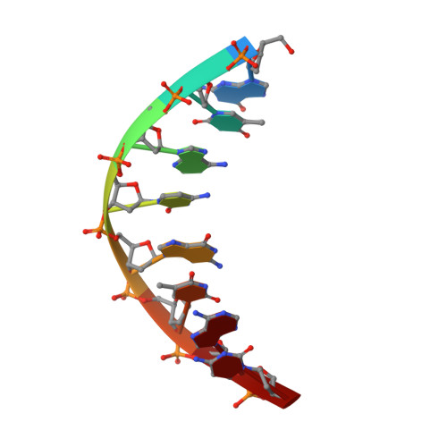
1D18, 1D19 - PubMed Abstract:
The solution structures of two alternating purine-pyrimidine octamers, [d(G-T-A-C-G-T-A-C)]2 and the reverse sequence [d(C-A-T-G-C-A-T-G)]2, are investigated by using nuclear magnetic resonance spectroscopy and restrained molecular dynamics calculations. Chemical shift assignments are obtained for non-exchangeable protons by a combination of two-dimensional correlation and nuclear Overhauser enhancement (NOE) spectroscopy experiments. Distances between protons are estimated by extrapolating distances derived from time-dependent NOE measurements to zero mixing time. Approximate dihedral angles are determined within the deoxyribose ring from coupling constants observed in one and two-dimensional spectra. Sets of distance and dihedral determinations for each of the duplexes form the bases for structure determination. Molecular dynamics is then used to generate structures that satisfy the experimental restraints incorporated as effective potentials into the total energy. Separate runs start from classical A and B-form DNA and converge to essentially identical structures. To circumvent the problems of spin diffusion and differential motion associated with distance measurements within molecules, models are improved by NOE-based refinement in which observed NOE intensities are compared to those calculated using a full matrix analysis procedure. The refined structures generally have the global features of B-type DNA. Some, but not all, variations in dihedral angles and in the spatial relationships of adjacent base-pairs are observed to be in synchrony with the alternating purine-pyrimidine sequence.
Organizational Affiliation:
Department of Biochemistry, University of Alberta, Edmonton, Canada.