1CYC

THE CRYSTAL STRUCTURE OF BONITO (KATSUO) FERROCYTOCHROME C AT 2.3 ANGSTROMS RESOLUTION. II. STRUCTURE AND FUNCTION


Experimental Data Snapshot

  • Method: X-RAY DIFFRACTION
  • Resolution: 2.30 Å

wwPDB Validation   3D Report Full Report


This is version 2.0 of the entry. See complete history


Literature

The crystal structure of bonito (katsuo) ferrocytochrome c at 2.3 A resolution. II. Structure and function.

Tanaka, N.Yamane, T.Tsukihara, T.Ashida, T.Kakudo, M.

(1975) J Biochem 77: 147-162

  • Primary Citation of Related Structures:  
    1CYC

  • PubMed Abstract: 

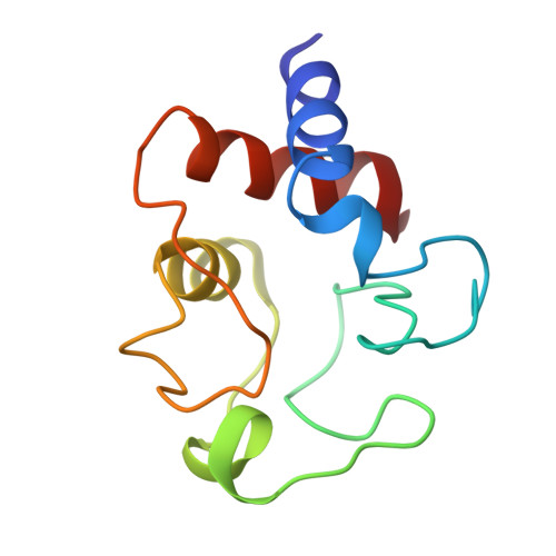
    The structure analysis of bonito heart ferrocytochrome c was carried out at 2.3 A resolution by X-ray diffraction, and a Kendrew-type skeletal model was built up. This molecule has an overall egg shape, 35 A in height, 30 A in width and 23 A in thickness; the 5th ligand of the heme iron atom is the N-epsilon atom of the His-18 imidazole ring and the 6th is the Met-80 sulfur atom. Distinct alpha-helix regions are found between the N-terminus and reside 11, between 60 and 69, and between 90 and the C-terminus. The most distinct difference between the conformation of the present molecule and that of the horse oxidized molecule is the location of the Phe-82 phenyl ring. In the present reduced molecule, the phyenyl ring is in closer contact with the iron atom and gives influences on the character of the iron atom. Inside the molecule, at the lower part of the heme pocket, there is an extended hydrogen bond network including the propionic acid residues of the heme group. Both Phe-82 and the hydrogen bond network may play a key role in the function of this molecule.


Macromolecules
Find similar proteins by:  (by identity cutoff)  |  3D Structure
Entity ID: 1
MoleculeChains Sequence LengthOrganismDetailsImage
FERROCYTOCHROME C
A, B
103Katsuwonus pelamisMutation(s): 0 
UniProt
Find proteins for P00025 (Katsuwonus pelamis)
Explore P00025 
Go to UniProtKB:  P00025
Entity Groups  
Sequence Clusters30% Identity50% Identity70% Identity90% Identity95% Identity100% Identity
UniProt GroupP00025
Sequence Annotations
Expand
  • Reference Sequence
Small Molecules
Ligands 1 Unique
IDChains Name / Formula / InChI Key2D Diagram3D Interactions
HEC
Query on HEC

Download Ideal Coordinates CCD File 
C [auth A],
D [auth B]
HEME C
C34 H34 Fe N4 O4
HXQIYSLZKNYNMH-LJNAALQVSA-N
Experimental Data & Validation

Experimental Data

  • Method: X-RAY DIFFRACTION
  • Resolution: 2.30 Å
  • Space Group: P 21 21 21
Unit Cell:
Length ( Å )Angle ( ˚ )
a = 57.68α = 90
b = 84.58β = 90
c = 37.83γ = 90

Structure Validation

View Full Validation Report


Entry History 

Deposition Data

Revision History  (Full details and data files)

  • Version 1.0: 1976-10-06
    Type: Initial release
  • Version 1.1: 2008-03-24
    Changes: Version format compliance
  • Version 1.2: 2011-07-13
    Changes: Version format compliance
  • Version 1.3: 2017-11-29
    Changes: Derived calculations, Other
  • Version 2.0: 2021-03-03
    Changes: Advisory, Atomic model, Data collection, Derived calculations, Non-polymer description, Structure summary