Context dependence of protein secondary structure formation: the three-dimensional structure and stability of a hybrid between chymotrypsin inhibitor 2 and helix E from subtilisin Carlsberg.
Osmark, P., Sorensen, P., Poulsen, F.M.(1993) Biochemistry 32: 11007-11014
- PubMed: 8218165
- DOI: https://doi.org/10.1021/bi00092a009
- Primary Citation of Related Structures:
1CIS - PubMed Abstract:
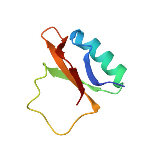
The loop region of chymotrypsin inhibitor 2 from barley has been employed as a scaffold for testing the intrinsic propensity of a peptide fragment to form a secondary structure. The helix formation of the nine amino acid residue segment Lys-Gln-Ala-Val-Asp-Asn-Ala-Tyr-Ala of helix E from subtilisin Carlsberg has been studied by the construction of a hybrid consisting of chymotrypsin inhibitor 2 (CI2) where part of the active loop has been replaced by the nonapeptide. An expression system for a truncated form of CI2 where the 19 structureless residues of the N-terminus have been removed and Leu20 replaced by methionyl was constructed from the entire 83-residue wild-type CI2 gene by polymerase chain reaction methodology. The gene encoding the hybrid was constructed from the truncated inhibitor gene. The stability of the truncated inhibitor and of the hybrid toward guanidinium chloride denaturation was examined. From these measurements, the energy of unfolding in pure water was extrapolated to 30.5 +/- 1.0 kJ/mol for the truncated inhibitor and 10.9 +/- 0.3 kJ/mol for the hybrid. These energies show that the stability of CI2 is unaffected by the N-terminal truncation but severely decreased by the loop mutations. The three-dimensional structure of the hybrid protein has been determined in solution by nuclear magnetic resonance spectroscopy using 893 distance restraints and 84 torsional angle restraints. The average root-mean-square deviation (rmsd) of 15 structures compared to their geometrical average was 0.8 +/- 0.2 A for heavy backbone atoms and 1.3 +/- 0.2 A for all heavy atoms.(ABSTRACT TRUNCATED AT 250 WORDS)
Organizational Affiliation:
Carlsberg Laboratorium, Kemisk Afdeling, Copenhagen, Denmark.