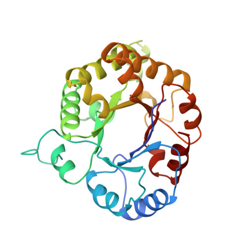
Crystal structure of triosephosphate isomerase from Trypanosoma cruzi in hexane.
Gao, X.G., Maldonado, E., Perez-Montfort, R., Garza-Ramos, G., de Gomez-Puyou, M.T., Gomez-Puyou, A., Rodriguez-Romero, A.(1999) Proc Natl Acad Sci U S A 96: 10062-10067
- PubMed: 10468562
- DOI: https://doi.org/10.1073/pnas.96.18.10062
- Primary Citation of Related Structures:
1CI1 - PubMed Abstract:
To gain insight into the mechanisms of enzyme catalysis in organic solvents, the x-ray structure of some monomeric enzymes in organic solvents was determined. However, it remained to be explored whether the structure of oligomeric proteins is also amenable to such analysis. The field acquired new perspectives when it was proposed that the x-ray structure of enzymes in nonaqueous media could reveal binding sites for organic solvents that in principle could represent the starting point for drug design. Here, a crystal of the dimeric enzyme triosephosphate isomerase from the pathogenic parasite Trypanosoma cruzi was soaked and diffracted in hexane and its structure solved at 2-A resolution. Its overall structure and the dimer interface were not altered by hexane. However, there were differences in the orientation of the side chains of several amino acids, including that of the catalytic Glu-168 in one of the monomers. No hexane molecules were detected in the active site or in the dimer interface. However, three hexane molecules were identified on the surface of the protein at sites, which in the native crystal did not have water molecules. The number of water molecules in the hexane structure was higher than in the native crystal. Two hexanes localized at <4 A from residues that form the dimer interface; they were in close proximity to a site that has been considered a potential target for drug design.
Organizational Affiliation:
Instituto de Fisiología Celular, Universidad Nacional Autónoma de México, 04510 México D. F., Mexico.