Membrane-binding peptide from the C2 domain of factor VIII forms an amphipathic structure as determined by NMR spectroscopy.
Gilbert, G.E., Baleja, J.D.(1995) Biochemistry 34: 3022-3031
- PubMed: 7893714
- DOI: https://doi.org/10.1021/bi00009a033
- Primary Citation of Related Structures:
1CFG - PubMed Abstract:
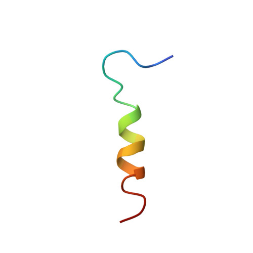
Factor VIII binds to cell membranes prior to assembling with the serine protease, factor IXa, to form the factor X-activating enzyme complex. In order to better understand the interaction between factor VIII and phosphatidylserine-containing membranes, we have synthesized the membrane-binding peptide from the C2 domain of factor VIII, corresponding to residues 2303-2324. The peptide, fVIII2303-24, with a primary structure of TRYLRIHPQSWVHQIALRMEVL, aggregates at concentrations above 2 microM at pH 7 but is soluble at pH 6. fVIII2303-24 competes with fluorescein-labeled factor VIII (Ki = 3 microM) for binding sites on synthetic phosphatidylserine-containing membranes and for binding sites on stimulated platelets. Circular dichroism spectra indicate that fVIII2303-24 is predominantly a random coil in aqueous solution but adopts a predominantly helical conformation upon interaction with SDS micelles. 1H NMR spectroscopy in the presence of SDS micelles allowed estimation of interproton distances from the nuclear Overhauser effect and estimation of torsion angles from coupling constants indicated by splitting of resonance lines. The distance and angle estimates, processed by distance geometry/simulated annealing software, indicate that fVIII2303-24 has an alpha-helical segment encompassing residues P8-E20 and an extended segment encompassing residues L4-P8. The location of six hydrophobic residues on one face of the structure suggests that hydrophobic interactions contribute to membrane-binding. In addition, two arginines penetrate the hydrophobic plane suggesting that they interact with phosphate moieties in a phospholipid bilayer.
Organizational Affiliation:
Medicine Department, Brockton-West Roxbury VA Medical Center, Boston, Massachusetts 02132.