Conformational flexibility of the acetylcholinesterase tetramer suggested by x-ray crystallography.
Bourne, Y., Grassi, J., Bougis, P.E., Marchot, P.(1999) J Biol Chem 274: 30370-30376
- PubMed: 10521413
- DOI: https://doi.org/10.1074/jbc.274.43.30370
- Primary Citation of Related Structures:
1C2B, 1C2O - PubMed Abstract:
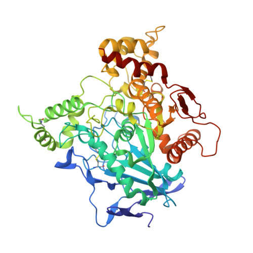
Acetylcholinesterase, a polymorphic enzyme, appears to form amphiphilic and nonamphiphilic tetramers from a single splice variant; this suggests discrete tetrameric arrangements where the amphipathic carboxyl-terminal sequences can be either buried or exposed. Two distinct, but related crystal structures of the soluble, trypsin-released tetramer of acetylcholinesterase from Electrophorus electricus were solved at 4.5 and 4.2 A resolution by molecular replacement. Resolution at these levels is sufficient to provide substantial information on the relative orientations of the subunits within the tetramer. The two structures, which show canonical homodimers of subunits assembled through four-helix bundles, reveal discrete geometries in the assembly of the dimers to form: (a) a loose, pseudo-square planar tetramer with antiparallel alignment of the two four-helix bundles and a large space in the center where the carboxyl-terminal sequences may be buried or (b) a compact, square nonplanar tetramer that may expose all four sequences on a single side. Comparison of these two structures points to significant conformational flexibility of the tetramer about the four-helix bundle axis and along the dimer-dimer interface. Hence, in solution, several conformational states of a flexible tetrameric arrangement of acetylcholinesterase catalytic subunits may exist to accommodate discrete carboxyl-terminal sequences of variable dimensions and amphipathicity.
Organizational Affiliation:
CNRS, Unité Propre de Recherche 9039, Architecture et Fonction des Macromolécules Biologiques, Institut de Biologie et Microbiologie Structurale, F-13402 Marseille Cedex 20, France.