1BCH

MANNOSE-BINDING PROTEIN-A MUTANT (QPDWGH) COMPLEXED WITH N-ACETYL-D-GALACTOSAMINE


Experimental Data Snapshot

  • Method: X-RAY DIFFRACTION
  • Resolution: 2.00 Å
  • R-Value Free: 0.252 
  • R-Value Work: 0.219 
  • R-Value Observed: 0.219 

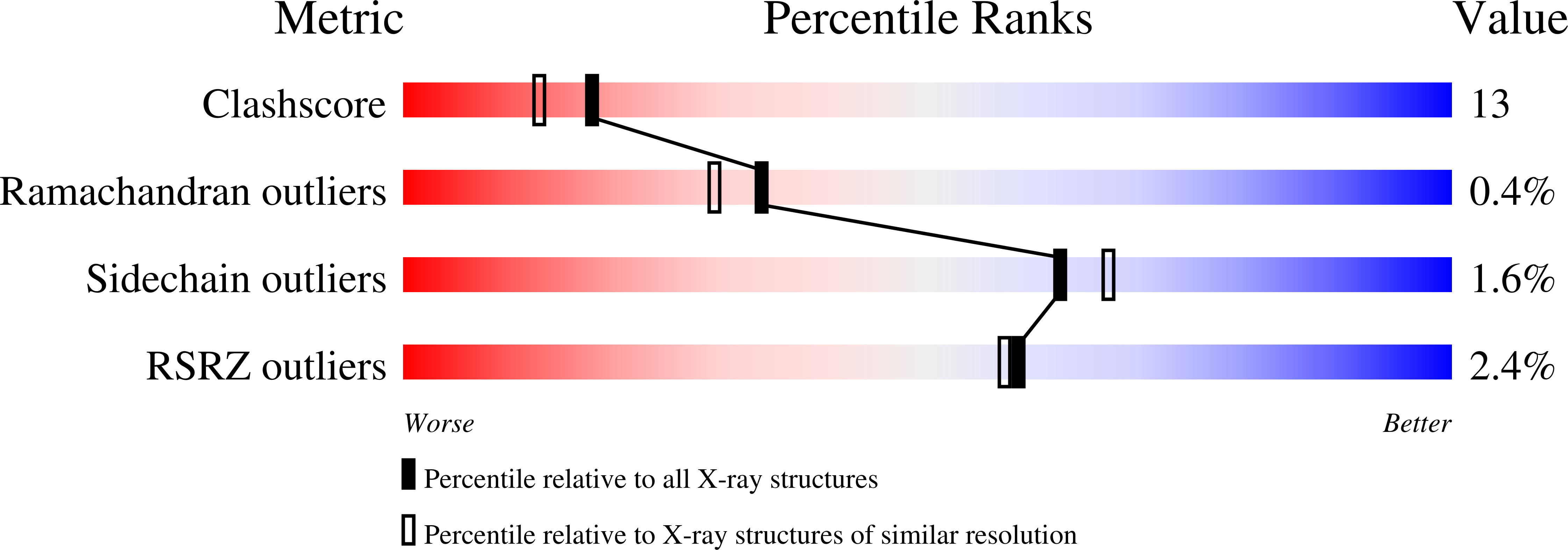
wwPDB Validation   3D Report Full Report


Ligand Structure Quality Assessment 


This is version 2.2 of the entry. See complete history


Literature

Mechanism of N-acetylgalactosamine binding to a C-type animal lectin carbohydrate-recognition domain.

Kolatkar, A.R.Leung, A.K.Isecke, R.Brossmer, R.Drickamer, K.Weis, W.I.

(1998) J Biol Chem 273: 19502-19508

  • DOI: https://doi.org/10.1074/jbc.273.31.19502
  • Primary Citation of Related Structures:  
    1BCH, 1BCJ

  • PubMed Abstract: 

    The mammalian hepatic asialoglycoprotein receptor, a member of the C-type animal lectin family, displays preferential binding to N-acetylgalactosamine compared with galactose. The structural basis for selective binding to N-acetylgalactosamine has been investigated. Regions of the carbohydrate-recognition domain of the receptor believed to be important in preferential binding to N-acetylgalactosamine have been inserted into the homologous carbohydrate-recognition domain of a mannose-binding protein mutant that was previously altered to bind galactose. Introduction of a single histidine residue corresponding to residue 256 of the hepatic asialoglycoprotein receptor was found to cause a 14-fold increase in the relative affinity for N-acetylgalactosamine compared with galactose. The relative ability of various acyl derivatives of galactosamine to compete for binding to this modified carbohydrate-recognition domain suggest that it is a good model for the natural N-acetylgalactosamine binding site of the asialoglycoprotein receptor. Crystallographic analysis of this mutant carbohydrate-recognition domain in complex with N-acetylgalactosamine reveals a direct interaction between the inserted histidine residue and the methyl group of the N-acetyl substituent of the sugar. Evidence for the role of the side chain at position 208 of the receptor in positioning this key histidine residue was obtained from structural analysis and mutagenesis experiments. The corresponding serine residue in the modified carbohydrate-recognition domain of mannose-binding protein forms a hydrogen bond to the imidazole side chain. When this serine residue is changed to valine, loss in selectivity for N-acetylgalactosamine is observed. The structure of this mutant reveals that the beta-branched valine side chain interacts directly with the histidine side chain, resulting in an altered imidazole ring orientation.


  • Organizational Affiliation

    Department of Structural Biology, Stanford University School of Medicine, Stanford, California 94305, USA.


Macromolecules
Find similar proteins by:  (by identity cutoff)  |  3D Structure
Entity ID: 1
MoleculeChains Sequence LengthOrganismDetailsImage
MANNOSE-BINDING PROTEIN-AA [auth 1],
B [auth 2],
C [auth 3]
154Rattus norvegicusMutation(s): 6 
UniProt
Find proteins for P19999 (Rattus norvegicus)
Explore P19999 
Go to UniProtKB:  P19999
Entity Groups  
Sequence Clusters30% Identity50% Identity70% Identity90% Identity95% Identity100% Identity
UniProt GroupP19999
Sequence Annotations
Expand
  • Reference Sequence
Small Molecules
Ligands 5 Unique
IDChains Name / Formula / InChI Key2D Diagram3D Interactions
NGA
Query on NGA

Download Ideal Coordinates CCD File 
I [auth 2],
O [auth 3]
2-acetamido-2-deoxy-beta-D-galactopyranose
C8 H15 N O6
OVRNDRQMDRJTHS-JAJWTYFOSA-N
A2G
Query on A2G

Download Ideal Coordinates CCD File 
D [auth 1]2-acetamido-2-deoxy-alpha-D-galactopyranose
C8 H15 N O6
OVRNDRQMDRJTHS-CBQIKETKSA-N
CA
Query on CA

Download Ideal Coordinates CCD File 
E [auth 1]
F [auth 1]
G [auth 1]
J [auth 2]
K [auth 2]
E [auth 1],
F [auth 1],
G [auth 1],
J [auth 2],
K [auth 2],
L [auth 2],
P [auth 3],
Q [auth 3],
R [auth 3]
CALCIUM ION
Ca
BHPQYMZQTOCNFJ-UHFFFAOYSA-N
CL
Query on CL

Download Ideal Coordinates CCD File 
H [auth 1],
M [auth 2],
S [auth 3]
CHLORIDE ION
Cl
VEXZGXHMUGYJMC-UHFFFAOYSA-M
NA
Query on NA

Download Ideal Coordinates CCD File 
N [auth 2]SODIUM ION
Na
FKNQFGJONOIPTF-UHFFFAOYSA-N
Experimental Data & Validation

Experimental Data

  • Method: X-RAY DIFFRACTION
  • Resolution: 2.00 Å
  • R-Value Free: 0.252 
  • R-Value Work: 0.219 
  • R-Value Observed: 0.219 
  • Space Group: C 1 2 1
Unit Cell:
Length ( Å )Angle ( ˚ )
a = 80.49α = 90
b = 85.01β = 104.82
c = 98.71γ = 90
Software Package:
Software NamePurpose
DENZOdata reduction
SCALEPACKdata scaling
CNSrefinement
CNSphasing

Structure Validation

View Full Validation Report


Ligand Structure Quality Assessment 


Entry History 

Deposition Data

Revision History  (Full details and data files)

  • Version 1.0: 1998-06-17
    Type: Initial release
  • Version 1.1: 2008-03-03
    Changes: Version format compliance
  • Version 1.2: 2011-07-13
    Changes: Version format compliance
  • Version 2.0: 2020-07-29
    Type: Remediation
    Reason: Carbohydrate remediation
    Changes: Advisory, Atomic model, Data collection, Database references, Derived calculations, Structure summary
  • Version 2.1: 2021-11-03
    Changes: Database references, Structure summary
  • Version 2.2: 2023-08-02
    Changes: Refinement description