1AA0

FIBRITIN DELETION MUTANT E (BACTERIOPHAGE T4)


Experimental Data Snapshot

  • Method: X-RAY DIFFRACTION
  • Resolution: 2.20 Å
  • R-Value Free: 0.259 
  • R-Value Work: 0.216 
  • R-Value Observed: 0.216 

wwPDB Validation   3D Report Full Report


This is version 1.4 of the entry. See complete history


Literature

Structure of bacteriophage T4 fibritin: a segmented coiled coil and the role of the C-terminal domain.

Tao, Y.Strelkov, S.V.Mesyanzhinov, V.V.Rossmann, M.G.

(1997) Structure 5: 789-798

  • DOI: https://doi.org/10.1016/s0969-2126(97)00233-5
  • Primary Citation of Related Structures:  
    1AA0

  • PubMed Abstract: 

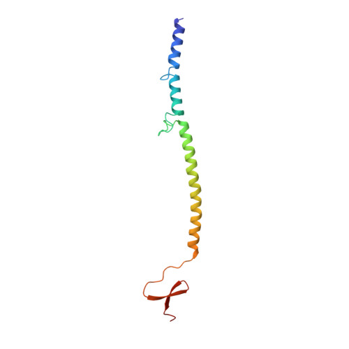
    Oligomeric coiled-coil motifs are found in numerous protein structures; among them is fibritin, a structural protein of bacteriophage T4, which belongs to a class of chaperones that catalyze a specific phage-assembly process. Fibritin promotes the assembly of the long tail fibers and their subsequent attachment to the tail baseplate; it is also a sensing device that controls the retraction of the long tail fibers in adverse environments and, thus, prevents infection. The structure of fibritin had been predicted from sequence and biochemical analyses to be mainly a triple-helical coiled coil. The determination of its structure at atomic resolution was expected to give insights into the assembly process and biological function of fibritin, and the properties of modified coiled-coil structures in general.


  • Organizational Affiliation

    Department of Biological Sciences, Purdue University, West Lafayette, Indiana 47907-1392, USA.


Macromolecules
Find similar proteins by:  (by identity cutoff)  |  3D Structure
Entity ID: 1
MoleculeChains Sequence LengthOrganismDetailsImage
FIBRITIN113Tequatrovirus T4Mutation(s): 0 
Gene Names: WAC
UniProt
Find proteins for P10104 (Enterobacteria phage T4)
Explore P10104 
Go to UniProtKB:  P10104
Entity Groups  
Sequence Clusters30% Identity50% Identity70% Identity90% Identity95% Identity100% Identity
UniProt GroupP10104
Sequence Annotations
Expand
  • Reference Sequence
Experimental Data & Validation

Experimental Data

  • Method: X-RAY DIFFRACTION
  • Resolution: 2.20 Å
  • R-Value Free: 0.259 
  • R-Value Work: 0.216 
  • R-Value Observed: 0.216 
  • Space Group: H 3 2
Unit Cell:
Length ( Å )Angle ( ˚ )
a = 41.2α = 90
b = 41.2β = 90
c = 358.7γ = 120
Software Package:
Software NamePurpose
X-PLORmodel building
X-PLORrefinement
DENZOdata reduction
SCALEPACKdata scaling
X-PLORphasing

Structure Validation

View Full Validation Report


Entry History 

Deposition Data

Revision History  (Full details and data files)

  • Version 1.0: 1997-07-23
    Type: Initial release
  • Version 1.1: 2008-03-24
    Changes: Version format compliance
  • Version 1.2: 2011-07-13
    Changes: Derived calculations, Version format compliance
  • Version 1.3: 2011-11-16
    Changes: Atomic model
  • Version 1.4: 2024-02-07
    Changes: Data collection, Database references, Derived calculations, Other