1A9J

APYRIMIDINIC DNA WITH BOUND WATER AT THE DAMAGED SITE, BETA FORM, NMR, 1 STRUCTURE

  • Classification: DNA
  • Mutation(s): No 

  • Deposited: 1998-04-06 Released: 1998-07-15 
  • Deposition Author(s): Beger, R.D., Bolton, P.H.

Experimental Data Snapshot

  • Method: SOLUTION NMR
  • Conformers Calculated: 100 
  • Conformers Submitted: 
  • Selection Criteria: AVERAGE OF 5 STRUCTURES THAT THE NOESY BACK CALCULATION AGREES WITH EXPERIMENTAL NOESY 

wwPDB Validation   3D Report Full Report


This is version 1.4 of the entry. See complete history


Literature

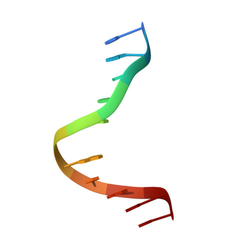
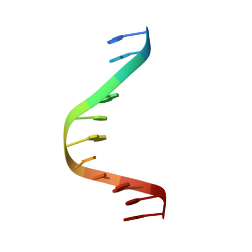
Structures of apurinic and apyrimidinic sites in duplex DNAs.

Beger, R.D.Bolton, P.H.

(1998) J Biol Chem 273: 15565-15573

  • DOI: https://doi.org/10.1074/jbc.273.25.15565
  • Primary Citation of Related Structures:  
    1A9G, 1A9H, 1A9I, 1A9J

  • PubMed Abstract: 

    Natural and exogenous processes can give rise to abasic sites with either a purine or pyrimidine as the base on the opposing strand. The solution state structures of the apyrimidinic DNA duplex, with D6 indicating an abasic site, [sequence: see text] referred to as AD, and the apurinic DNA duplex with a dC17, referred to as CD, have been determined. A particularly striking difference is that the abasic site in CD is predominantly a beta hemiacetal, whereas in AD the alpha and beta forms are equally present. Hydrogen bonding with water by the abasic site and the base on the opposite strand appears to play a large role in determining the structure near the damaged site. Comparison of these structures with that of a duplex DNA containing a thymine glycol at the same position as the abasic site and with that of a duplex DNA containing an abasic site in the middle of a curved DNA sequence offers some insight into the common and distinct structural features of damaged DNA sites.


  • Organizational Affiliation

    Department of Chemistry, Wesleyan University, Middletown, Connecticut 06459, USA.


Macromolecules

Find similar nucleic acids by:  Sequence   |   3D Structure  

Entity ID: 1
MoleculeChains LengthOrganismImage
DNA (5'-D(*CP*GP*CP*GP*AP*AABP*AP*CP*GP*CP*C)-3')11N/A
Sequence Annotations
Expand
  • Reference Sequence

Find similar nucleic acids by:  Sequence   |   3D Structure  

Entity ID: 2
MoleculeChains LengthOrganismImage
DNA (5'-D(*GP*GP*CP*GP*TP*AP*TP*CP*GP*CP*G)-3')11N/A
Sequence Annotations
Expand
  • Reference Sequence
Experimental Data & Validation

Experimental Data

  • Method: SOLUTION NMR
  • Conformers Calculated: 100 
  • Conformers Submitted: 
  • Selection Criteria: AVERAGE OF 5 STRUCTURES THAT THE NOESY BACK CALCULATION AGREES WITH EXPERIMENTAL NOESY 

Structure Validation

View Full Validation Report


Entry History 

Deposition Data

Revision History  (Full details and data files)

  • Version 1.0: 1998-07-15
    Type: Initial release
  • Version 1.1: 2008-03-24
    Changes: Version format compliance
  • Version 1.2: 2011-07-13
    Changes: Version format compliance
  • Version 1.3: 2014-07-09
    Changes: Structure summary
  • Version 1.4: 2024-04-10
    Changes: Data collection, Database references, Derived calculations, Other