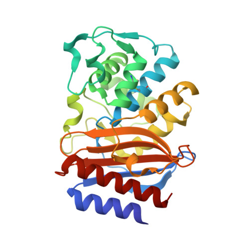
8DDZ

TEM-1 beta-lactamase A237Y


Experimental Data Snapshot

  • Method: X-RAY DIFFRACTION
  • Resolution: 1.45 Å
  • R-Value Free: 0.212 
  • R-Value Work: 0.177 
  • R-Value Observed: 0.179 

wwPDB Validation   3D Report Full Report


This is version 1.2 of the entry. See complete history


Literature

Protein Electric Fields Enable Faster and Longer-Lasting Covalent Inhibition of beta-Lactamases.

Ji, Z.Kozuch, J.Mathews, I.I.Diercks, C.S.Shamsudin, Y.Schulz, M.A.Boxer, S.G.

(2022) J Am Chem Soc 144: 20947-20954

  • DOI: https://doi.org/10.1021/jacs.2c09876
  • Primary Citation of Related Structures:  
    7U6Q, 8DDZ, 8DE0, 8DE1, 8DE2

  • PubMed Abstract: 

    The widespread design of covalent drugs has focused on crafting reactive groups of proper electrophilicity and positioning toward targeted amino-acid nucleophiles. We found that environmental electric fields projected onto a reactive chemical bond, an overlooked design element, play essential roles in the covalent inhibition of TEM-1 β-lactamase by avibactam. Using the vibrational Stark effect, the magnitudes of the electric fields that are exerted by TEM active sites onto avibactam's reactive C═O were measured and demonstrate an electrostatic gating effect that promotes bond formation yet relatively suppresses the reverse dissociation. These results suggest new principles of covalent drug design and off-target site prediction. Unlike shape and electrostatic complementary which address binding constants, electrostatic catalysis drives reaction rates, essential for covalent inhibition, and deepens our understanding of chemical reactivity, selectivity, and stability in complex systems.


  • Organizational Affiliation

    Department of Chemistry, Stanford University, Stanford, California 94305, United States.


Macromolecules
Find similar proteins by:  (by identity cutoff)  |  3D Structure
Entity ID: 1
MoleculeChains Sequence LengthOrganismDetailsImage
Beta-lactamase TEM
A, B, C, D
264Escherichia coliMutation(s): 2 
Gene Names: blaTEM-1
EC: 3.5.2.6
UniProt
Find proteins for P62593 (Escherichia coli)
Explore P62593 
Go to UniProtKB:  P62593
Entity Groups  
Sequence Clusters30% Identity50% Identity70% Identity90% Identity95% Identity100% Identity
UniProt GroupP62593
Sequence Annotations
Expand
  • Reference Sequence
Experimental Data & Validation

Experimental Data

  • Method: X-RAY DIFFRACTION
  • Resolution: 1.45 Å
  • R-Value Free: 0.212 
  • R-Value Work: 0.177 
  • R-Value Observed: 0.179 
  • Space Group: P 1 21 1
Unit Cell:
Length ( Å )Angle ( ˚ )
a = 60.61α = 90
b = 83.77β = 90.23
c = 95.66γ = 90
Software Package:
Software NamePurpose
PHENIXrefinement
XSCALEdata scaling
PDB_EXTRACTdata extraction
XDSdata reduction
MOLREPphasing

Structure Validation

View Full Validation Report


Entry History & Funding Information

Deposition Data


Funding OrganizationLocationGrant Number
National Institutes of Health/National Institute of General Medical Sciences (NIH/NIGMS)United StatesGM118044
National Institutes of Health/National Institute of General Medical Sciences (NIH/NIGMS)United StatesP30GM133894

Revision History  (Full details and data files)

  • Version 1.0: 2022-09-07
    Type: Initial release
  • Version 1.1: 2023-03-22
    Changes: Database references
  • Version 1.2: 2023-10-25
    Changes: Data collection, Refinement description