8BJ4

Crystal structure of Medicago truncatula histidinol-phosphate aminotransferase (HISN6) in apo form


Experimental Data Snapshot

  • Method: X-RAY DIFFRACTION
  • Resolution: 1.45 Å
  • R-Value Free: 0.181 
  • R-Value Work: 0.148 
  • R-Value Observed: 0.148 

wwPDB Validation   3D Report Full Report


Ligand Structure Quality Assessment 


This is version 1.0 of the entry. See complete history


Literature

Insights into the substrate specificity, structure, and dynamics of plant histidinol-phosphate aminotransferase (HISN6).

Rutkiewicz, M.Nogues, I.Witek, W.Angelaccio, S.Contestabile, R.Ruszkowski, M.

(2023) Plant Physiol Biochem 196: 759-773

  • DOI: https://doi.org/10.1016/j.plaphy.2023.02.017
  • Primary Citation of Related Structures:  
    8BJ1, 8BJ2, 8BJ3, 8BJ4

  • PubMed Abstract: 

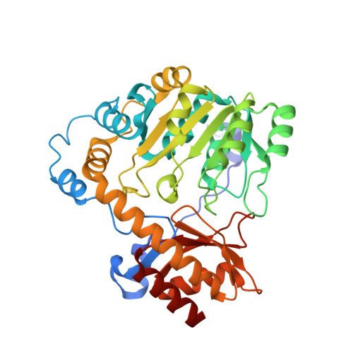
    Histidinol-phosphate aminotransferase is the sixth protein (hence HISN6) in the histidine biosynthetic pathway in plants. HISN6 is a pyridoxal 5'-phosphate (PLP)-dependent enzyme that catalyzes the reversible conversion of imidazole acetol phosphate into L-histidinol phosphate (HOLP). Here, we show that plant HISN6 enzymes are closely related to the orthologs from Chloroflexota. The studied example, HISN6 from Medicago truncatula (MtHISN6), exhibits a surprisingly high affinity for HOLP, which is much higher than reported for bacterial homologs. Moreover, unlike the latter, MtHISN6 does not transaminate phenylalanine. High-resolution crystal structures of MtHISN6 in the open and closed states, as well as the complex with HOLP and the apo structure without PLP, bring new insights into the enzyme dynamics, pointing at a particular role of a string-like fragment that oscillates near the active site and participates in the HOLP binding. When MtHISN6 is compared to bacterial orthologs with known structures, significant differences arise in or near the string region. The high affinity of MtHISN6 appears linked to the particularly tight active site cavity. Finally, a virtual screening against a library of over 1.3 mln compounds revealed three sites in the MtHISN6 structure with the potential to bind small molecules. Such compounds could be developed into herbicides inhibiting plant HISN6 enzymes absent in animals, which makes them a potential target for weed control agents.


  • Organizational Affiliation

    Department of Structural Biology of Eukaryotes, Institute of Bioorganic Chemistry, Polish Academy of Sciences, Poznan, Poland.


Macromolecules
Find similar proteins by:  (by identity cutoff)  |  3D Structure
Entity ID: 1
MoleculeChains Sequence LengthOrganismDetailsImage
histidinol-phosphate aminotransferase
A, B, C, D
363Medicago truncatulaMutation(s): 0 
Gene Names: 25495946MTR_6g033540
EC: 2.6.1.9
UniProt
Find proteins for A0A072U7F9 (Medicago truncatula)
Explore A0A072U7F9 
Go to UniProtKB:  A0A072U7F9
Entity Groups  
Sequence Clusters30% Identity50% Identity70% Identity90% Identity95% Identity100% Identity
UniProt GroupA0A072U7F9
Sequence Annotations
Expand
  • Reference Sequence
Small Molecules
Ligands 3 Unique
IDChains Name / Formula / InChI Key2D Diagram3D Interactions
SO4 (Subject of Investigation/LOI)
Query on SO4

Download Ideal Coordinates CCD File 
AA [auth C]
E [auth A]
EA [auth C]
FA [auth C]
GA [auth C]
AA [auth C],
E [auth A],
EA [auth C],
FA [auth C],
GA [auth C],
HA [auth C],
IA [auth C],
JA [auth C],
L [auth A],
O [auth A],
P [auth A],
SA [auth D],
TA [auth D],
U [auth B],
UA [auth D],
V [auth B],
W [auth B]
SULFATE ION
O4 S
QAOWNCQODCNURD-UHFFFAOYSA-L
EDO
Query on EDO

Download Ideal Coordinates CCD File 
BA [auth C]
CA [auth C]
DA [auth C]
F [auth A]
G [auth A]
BA [auth C],
CA [auth C],
DA [auth C],
F [auth A],
G [auth A],
H [auth A],
I [auth A],
J [auth A],
K [auth A],
LA [auth D],
M [auth A],
MA [auth D],
N [auth A],
NA [auth D],
OA [auth D],
PA [auth D],
QA [auth D],
R [auth B],
RA [auth D],
S [auth B],
T [auth B],
Y [auth C],
Z [auth C]
1,2-ETHANEDIOL
C2 H6 O2
LYCAIKOWRPUZTN-UHFFFAOYSA-N
NA
Query on NA

Download Ideal Coordinates CCD File 
KA [auth C],
Q [auth A],
VA [auth D],
X [auth B]
SODIUM ION
Na
FKNQFGJONOIPTF-UHFFFAOYSA-N
Experimental Data & Validation

Experimental Data

  • Method: X-RAY DIFFRACTION
  • Resolution: 1.45 Å
  • R-Value Free: 0.181 
  • R-Value Work: 0.148 
  • R-Value Observed: 0.148 
  • Space Group: P 1 21 1
Unit Cell:
Length ( Å )Angle ( ˚ )
a = 74.039α = 90
b = 93.082β = 106.57
c = 110.518γ = 90
Software Package:
Software NamePurpose
PHENIXrefinement
XSCALEdata scaling
PDB_EXTRACTdata extraction
XDSdata reduction
PHENIXphasing

Structure Validation

View Full Validation Report


Ligand Structure Quality Assessment 


Entry History & Funding Information

Deposition Data


Funding OrganizationLocationGrant Number
Polish National Science CentrePoland2018/31/D/NZ1/03630

Revision History  (Full details and data files)

  • Version 1.0: 2023-03-22
    Type: Initial release