7ZSC

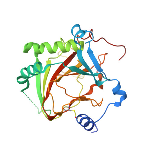
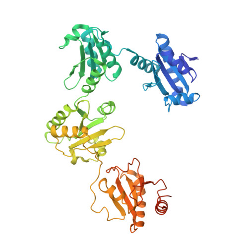
Crystal structure of the heterodimeric human C-P4H-II with truncated alpha subunit (C-P4H-II delta281)


Experimental Data Snapshot

  • Method: X-RAY DIFFRACTION
  • Resolution: 3.85 Å
  • R-Value Free: 0.278 
  • R-Value Work: 0.244 
  • R-Value Observed: 0.247 

wwPDB Validation   3D Report Full Report


This is version 1.2 of the entry. See complete history


Literature

Crystal structure of the collagen prolyl 4-hydroxylase (C-P4H) catalytic domain complexed with PDI: Toward a model of the C-P4H alpha 2 beta 2 tetramer.

Murthy, A.V.Sulu, R.Lebedev, A.Salo, A.M.Korhonen, K.Venkatesan, R.Tu, H.Bergmann, U.Janis, J.Laitaoja, M.Ruddock, L.W.Myllyharju, J.Koski, M.K.Wierenga, R.K.

(2022) J Biol Chem 298: 102614-102614

  • DOI: https://doi.org/10.1016/j.jbc.2022.102614
  • Primary Citation of Related Structures:  
    7ZSC

  • PubMed Abstract: 

    Collagen prolyl 4-hydroxylases (C-P4H) are α 2 β 2 tetramers, which catalyze the prolyl 4-hydroxylation of procollagen, allowing for the formation of the stable triple-helical collagen structure in the endoplasmic reticulum. The C-P4H α-subunit provides the N-terminal dimerization domain, the middle peptide-substrate-binding (PSB) domain, and the C-terminal catalytic (CAT) domain, whereas the β-subunit is identical to the enzyme protein disulfide isomerase (PDI). The structure of the N-terminal part of the α-subunit (N-terminal region and PSB domain) is known, but the structures of the PSB-CAT linker region and the CAT domain as well as its mode of assembly with the β/PDI subunit, are unknown. Here, we report the crystal structure of the CAT domain of human C-P4H-II complexed with the intact β/PDI subunit, at 3.8 Å resolution. The CAT domain interacts with the a, b', and a' domains of the β/PDI subunit, such that the CAT active site is facing bulk solvent. The structure also shows that the C-P4H-II CAT domain has a unique N-terminal extension, consisting of α-helices and a β-strand, which is the edge strand of its major antiparallel β-sheet. This extra region of the CAT domain interacts tightly with the β/PDI subunit, showing that the CAT-PDI interface includes an intersubunit disulfide bridge with the a' domain and tight hydrophobic interactions with the b' domain. Using this new information, the structure of the mature C-P4H-II α 2 β 2 tetramer is predicted. The model suggests that the CAT active-site properties are modulated by α-helices of the N-terminal dimerization domains of both subunits of the α 2 -dimer.


  • Organizational Affiliation

    Faculty of Biochemistry and Molecular Medicine, University of Oulu, Oulu, Finland; Biocenter Oulu, University of Oulu, Oulu, Finland.


Macromolecules
Find similar proteins by:  (by identity cutoff)  |  3D Structure
Entity ID: 1
MoleculeChains Sequence LengthOrganismDetailsImage
Prolyl 4-hydroxylase subunit alpha-2
A, B
262Homo sapiensMutation(s): 0 
Gene Names: P4HA2UNQ290/PRO330
EC: 1.14.11.2
UniProt & NIH Common Fund Data Resources
Find proteins for O15460 (Homo sapiens)
Explore O15460 
Go to UniProtKB:  O15460
PHAROS:  O15460
GTEx:  ENSG00000072682 
Entity Groups  
Sequence Clusters30% Identity50% Identity70% Identity90% Identity95% Identity100% Identity
UniProt GroupO15460
Sequence Annotations
Expand
  • Reference Sequence
Find similar proteins by:  (by identity cutoff)  |  3D Structure
Entity ID: 2
MoleculeChains Sequence LengthOrganismDetailsImage
Protein disulfide-isomerase
C, D
492Homo sapiensMutation(s): 0 
Gene Names: P4HBERBA2LPDIPDIA1PO4DB
EC: 5.3.4.1
UniProt & NIH Common Fund Data Resources
Find proteins for P07237 (Homo sapiens)
Explore P07237 
Go to UniProtKB:  P07237
PHAROS:  P07237
GTEx:  ENSG00000185624 
Entity Groups  
Sequence Clusters30% Identity50% Identity70% Identity90% Identity95% Identity100% Identity
UniProt GroupP07237
Sequence Annotations
Expand
  • Reference Sequence
Experimental Data & Validation

Experimental Data

  • Method: X-RAY DIFFRACTION
  • Resolution: 3.85 Å
  • R-Value Free: 0.278 
  • R-Value Work: 0.244 
  • R-Value Observed: 0.247 
  • Space Group: H 3
Unit Cell:
Length ( Å )Angle ( ˚ )
a = 252.879α = 90
b = 252.879β = 90
c = 89.4γ = 120
Software Package:
Software NamePurpose
BUSTERrefinement
XDSdata reduction
Aimlessdata scaling
MOLREPphasing
PHASERphasing

Structure Validation

View Full Validation Report


Entry History & Funding Information

Deposition Data


Funding OrganizationLocationGrant Number
Sigrid Juselius FoundationFinland--

Revision History  (Full details and data files)

  • Version 1.0: 2022-11-09
    Type: Initial release
  • Version 1.1: 2022-11-30
    Changes: Database references
  • Version 1.2: 2024-01-31
    Changes: Data collection, Refinement description