


CRYSTAL STRUCTURE OF THE P1 trifluoroethylglycine (TfeGly) BPTI MUTANT- BOVINE CHYMOTRYPSIN COMPLEX
Leppkes, J., Wehrhan, L., Dimos, N., Loll, B., Hohmann, T., Dyrks, M., Wieseke, A., Wahl, M.C., Keller, B.G., Koksch, B.To be published.
Experimental Data Snapshot
wwPDB Validation 3D Report Full Report

Entity ID: 1 | |||||
|---|---|---|---|---|---|
| Molecule | Chains | Sequence Length | Organism | Details | Image |
| Acetaldehyde dehydrogenase | 298 | Geobacillus stearothermophilus | Mutation(s): 0 Gene Names: pheF EC: 1.2.1.10 | 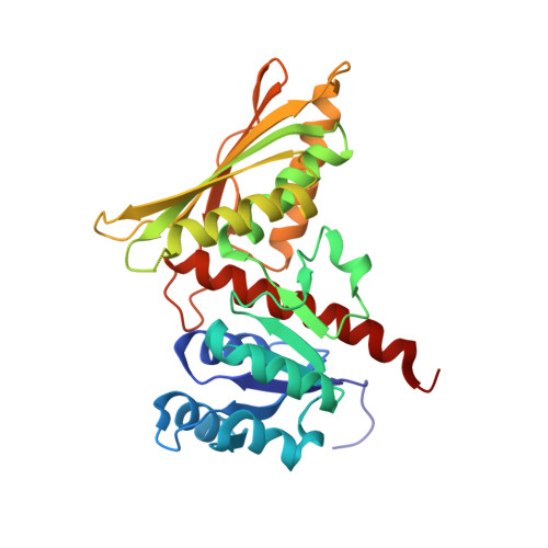 | |
UniProt | |||||
Find proteins for B0VXM6 (Geobacillus stearothermophilus) Explore B0VXM6 Go to UniProtKB: B0VXM6 | |||||
Entity Groups | |||||
| Sequence Clusters | 30% Identity50% Identity70% Identity90% Identity95% Identity100% Identity | ||||
| UniProt Group | B0VXM6 | ||||
Sequence AnnotationsExpand | |||||
| |||||
| Ligands 1 Unique | |||||
|---|---|---|---|---|---|
| ID | Chains | Name / Formula / InChI Key | 2D Diagram | 3D Interactions | |
| PO4 Query on PO4 | B [auth A], C [auth A], D [auth A] | PHOSPHATE ION O4 P NBIIXXVUZAFLBC-UHFFFAOYSA-K |  | ||
| Length ( Å ) | Angle ( ˚ ) |
|---|---|
| a = 69.25 | α = 90 |
| b = 69.25 | β = 90 |
| c = 88.4 | γ = 90 |
| Software Name | Purpose |
|---|---|
| PHENIX | refinement |
| XDS | data reduction |
| XSCALE | data scaling |
| PHASER | phasing |
| Funding Organization | Location | Grant Number |
|---|---|---|
| Not funded | -- |
RCSB PDB (citation) is hosted by
RCSB PDB is a member of the

