


crystal structure of Csn-PD from Paenibacillus dendritiformis
Sun, H.H., Cheng, Y.M., Cao, R., Liu, Q., Zhao, L.To be published.
Experimental Data Snapshot
wwPDB Validation 3D Report Full Report

Entity ID: 1 | |||||
|---|---|---|---|---|---|
| Molecule | Chains | Sequence Length | Organism | Details | Image |
| Chitosanase | 308 | Paenibacillus dendritiformis | Mutation(s): 0 Gene Names: DOE73_06680 EC: 3.2.1.132 | 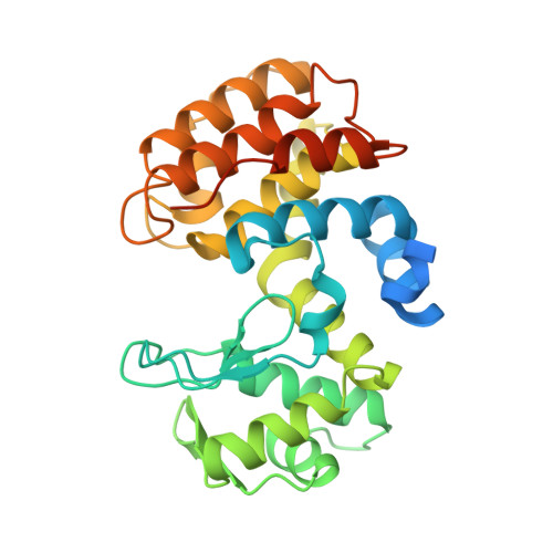 | |
UniProt | |||||
Find proteins for H3SPM3 (Paenibacillus dendritiformis C454) Explore H3SPM3 Go to UniProtKB: H3SPM3 | |||||
Entity Groups | |||||
| Sequence Clusters | 30% Identity50% Identity70% Identity90% Identity95% Identity100% Identity | ||||
| UniProt Group | H3SPM3 | ||||
Sequence AnnotationsExpand | |||||
| |||||
| Ligands 2 Unique | |||||
|---|---|---|---|---|---|
| ID | Chains | Name / Formula / InChI Key | 2D Diagram | 3D Interactions | |
| FLC Query on FLC | B [auth A] | CITRATE ANION C6 H5 O7 KRKNYBCHXYNGOX-UHFFFAOYSA-K |  | ||
| EDO Query on EDO | C [auth A] | 1,2-ETHANEDIOL C2 H6 O2 LYCAIKOWRPUZTN-UHFFFAOYSA-N |  | ||
| Length ( Å ) | Angle ( ˚ ) |
|---|---|
| a = 68.51 | α = 90 |
| b = 73.138 | β = 90 |
| c = 99.975 | γ = 90 |
| Software Name | Purpose |
|---|---|
| REFMAC | refinement |
| XDS | data reduction |
| Aimless | data scaling |
| PHASER | phasing |
| Funding Organization | Location | Grant Number |
|---|---|---|
| National Natural Science Foundation of China (NSFC) | China | 31801574 |
RCSB PDB (citation) is hosted by
RCSB PDB is a member of the

