7T3U

IP3, ATP, and Ca2+ bound type 3 IP3 receptor in the inactive state


Experimental Data Snapshot

  • Method: ELECTRON MICROSCOPY
  • Resolution: 3.70 Å
  • Aggregation State: PARTICLE 
  • Reconstruction Method: SINGLE PARTICLE 

wwPDB Validation   3D Report Full Report


This is version 1.1 of the entry. See complete history


Literature

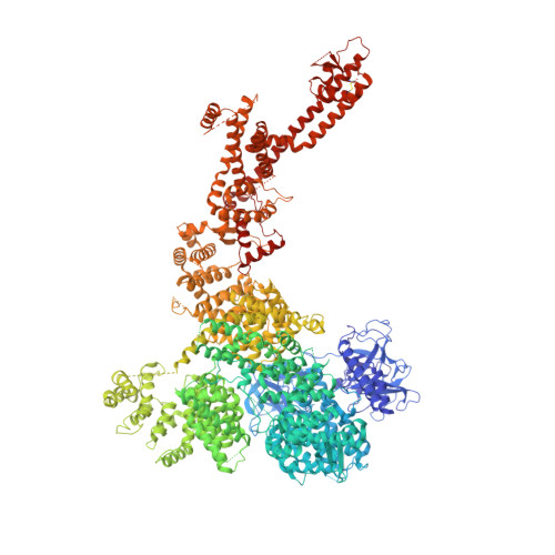
Structural basis for activation and gating of IP 3 receptors.

Schmitz, E.A.Takahashi, H.Karakas, E.

(2022) Nat Commun 13: 1408-1408

  • DOI: https://doi.org/10.1038/s41467-022-29073-2
  • Primary Citation of Related Structures:  
    7T3P, 7T3Q, 7T3R, 7T3T, 7T3U

  • PubMed Abstract: 

    A pivotal component of the calcium (Ca 2+ ) signaling toolbox in cells is the inositol 1,4,5-triphosphate (IP 3 ) receptor (IP 3 R), which mediates Ca 2+ release from the endoplasmic reticulum (ER), controlling cytoplasmic and organellar Ca 2+ concentrations. IP 3 Rs are co-activated by IP 3 and Ca 2+ , inhibited by Ca 2+ at high concentrations, and potentiated by ATP. However, the underlying molecular mechanisms are unclear. Here we report cryo-electron microscopy (cryo-EM) structures of human type-3 IP 3 R obtained from a single dataset in multiple gating conformations: IP 3 -ATP bound pre-active states with closed channels, IP 3 -ATP-Ca 2+ bound active state with an open channel, and IP 3 -ATP-Ca 2+ bound inactive state with a closed channel. The structures demonstrate how IP 3 -induced conformational changes prime the receptor for activation by Ca 2+ , how Ca 2+ binding leads to channel opening, and how ATP modulates the activity, providing insights into the long-sought questions regarding the molecular mechanism underpinning receptor activation and gating.


  • Organizational Affiliation

    Department of Molecular Physiology and Biophysics, Vanderbilt University, School of Medicine, Nashville, TN, 37232, USA.


Macromolecules
Find similar proteins by:  (by identity cutoff)  |  3D Structure
Entity ID: 1
MoleculeChains Sequence LengthOrganismDetailsImage
Inositol 1,4,5-trisphosphate receptor type 3
A, B, C, D
2,633Homo sapiensMutation(s): 0 
Gene Names: ITPR3
Membrane Entity: Yes 
UniProt & NIH Common Fund Data Resources
Find proteins for Q14573 (Homo sapiens)
Explore Q14573 
Go to UniProtKB:  Q14573
PHAROS:  Q14573
GTEx:  ENSG00000096433 
Entity Groups  
Sequence Clusters30% Identity50% Identity70% Identity90% Identity95% Identity100% Identity
UniProt GroupQ14573
Sequence Annotations
Expand
  • Reference Sequence
Small Molecules
Ligands 4 Unique
IDChains Name / Formula / InChI Key2D Diagram3D Interactions
ATP (Subject of Investigation/LOI)
Query on ATP

Download Ideal Coordinates CCD File 
G [auth A],
J [auth B],
M [auth C],
Q [auth D]
ADENOSINE-5'-TRIPHOSPHATE
C10 H16 N5 O13 P3
ZKHQWZAMYRWXGA-KQYNXXCUSA-N
I3P (Subject of Investigation/LOI)
Query on I3P

Download Ideal Coordinates CCD File 
F [auth A],
P [auth D]
D-MYO-INOSITOL-1,4,5-TRIPHOSPHATE
C6 H15 O15 P3
MMWCIQZXVOZEGG-XJTPDSDZSA-N
ZN (Subject of Investigation/LOI)
Query on ZN

Download Ideal Coordinates CCD File 
E [auth A],
I [auth B],
L [auth C],
O [auth D]
ZINC ION
Zn
PTFCDOFLOPIGGS-UHFFFAOYSA-N
CA (Subject of Investigation/LOI)
Query on CA

Download Ideal Coordinates CCD File 
H [auth A],
K [auth B],
N [auth C],
R [auth D]
CALCIUM ION
Ca
BHPQYMZQTOCNFJ-UHFFFAOYSA-N
Experimental Data & Validation

Experimental Data

  • Method: ELECTRON MICROSCOPY
  • Resolution: 3.70 Å
  • Aggregation State: PARTICLE 
  • Reconstruction Method: SINGLE PARTICLE 
EM Software:
TaskSoftware PackageVersion
MODEL REFINEMENTPHENIX
RECONSTRUCTIONcryoSPARC

Structure Validation

View Full Validation Report


Entry History & Funding Information

Deposition Data


Funding OrganizationLocationGrant Number
National Institutes of Health/National Institute of General Medical Sciences (NIH/NIGMS)United StatesR01GM141251
National Institutes of Health/National Institute of Diabetes and Digestive and Kidney Disease (NIH/NIDDK)United StatesP30DK020593
National Institutes of Health/National Institute of General Medical Sciences (NIH/NIGMS)United StatesT32GM008320

Revision History  (Full details and data files)

  • Version 1.0: 2022-03-23
    Type: Initial release
  • Version 1.1: 2022-05-04
    Changes: Database references