Structural and Biochemical Characterization of the Human Angiogenin-Proliferating Cell Nuclear Antigen Interaction.
Papaioannou, O.S.E., Tsika, A.C., Rovoli, M., Papadopoulos, G.E., Kontopidis, G., Spyroulias, G.A., Leonidas, D.D.(2023) Biochemistry 62: 1706-1715
- PubMed: 37218877
- DOI: https://doi.org/10.1021/acs.biochem.3c00158
- Primary Citation of Related Structures:
7PNJ, 7PNP, 7PNR - PubMed Abstract:
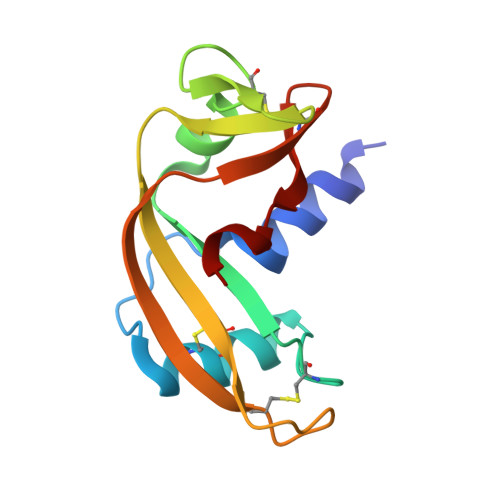
The molecular details of the interaction between human angiogenin (hAng) and proliferating cell nuclear antigen (PCNA) have been investigated by isothermal titration calorimetry (ITC), mutagenesis, and NMR spectroscopy. The two proteins were shown to interact directly through immunoprecipitation studies of hAng with PCNA in vitro , and their interaction was quantified by ITC, obtaining information on stoichiometry, enthalpy, entropy, and binding kinetics of the association. The hAng-PCNA association is strong, with a K d value of 126 nM. The interaction surface was mapped by NMR spectroscopy, indicating participating residues. A structural model for the PCNA-hAng complex was constructed by docking and molecular dynamics simulations based on NMR data. The model was validated by mutating the hAng residues Arg5 and Arg101, which seem critical for the complex formation, to glutamate. ITC experiments showed that the angiogenin variants R5E and R5ER101E displayed 6.5 and 7.8 times higher K d values, respectively, than that of the native protein, indicating the correctness of the model. The hAng S28AT36AS37A and hAng S28AT36AS37AS87A variants were also tested as positive controls, further supporting the validity of the model. The crystal structures of the hAng variants S28AT36AS37A and S28AT36AS37AS87A showed that the mutations did not cause any significant conformational change. This study presents evidence for the structural mode of the hAng-PCNA interaction, revealing valuable information about the angiogenin and PCNA biological roles in the cytoplasm.
Organizational Affiliation:
Department of Biochemistry and Biotechnology, University of Thessaly, Biopolis, 41500Larissa, Greece.