7O3K

Structure of a monomeric variant (L135E) of Sandercyanin fluorescent protein bound to biliverdin IX-alpha


Experimental Data Snapshot

  • Method: X-RAY DIFFRACTION
  • Resolution: 2.75 Å
  • R-Value Free: 0.251 
  • R-Value Work: 0.231 
  • R-Value Observed: 0.232 

wwPDB Validation   3D Report Full Report


This is version 1.2 of the entry. See complete history


Literature

Modulation of biliverdin dynamics and spectral properties by Sandercyanin.

Ghosh, S.Mondal, S.Yadav, K.Aggarwal, S.Schaefer, W.F.Narayana, C.Subramanian, R.

(2022) RSC Adv 12: 20296-20304

  • DOI: https://doi.org/10.1039/d2ra02880h
  • Primary Citation of Related Structures:  
    7O2Y, 7O3K, 7YX1

  • PubMed Abstract: 

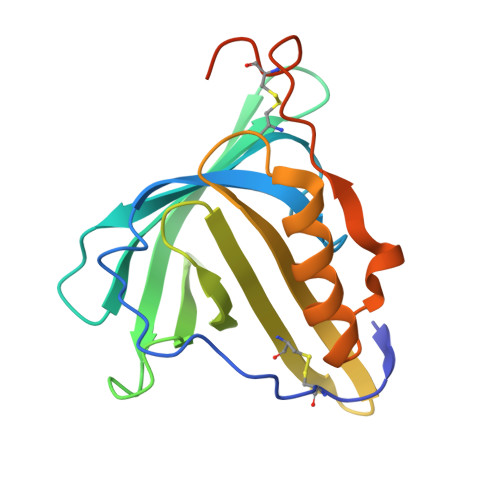
    Biliverdin IX-alpha (BV), a tetrapyrrole, is found ubiquitously in most living organisms. It functions as a metabolite, pigment, and signaling compound. While BV is known to bind to diverse protein families such as heme-metabolizing enzymes and phytochromes, not many BV-bound lipocalins (ubiquitous, small lipid-binding proteins) have been studied. The molecular basis of binding and conformational selectivity of BV in lipocalins remains unexplained. Sandercyanin (SFP)-BV complex is a blue lipocalin protein present in the mucus of the Canadian walleye ( Stizostedion vitreum ). In this study, we present the structures and binding modes of BV to SFP. Using a combination of designed site-directed mutations, X-ray crystallography, UV/VIS, and resonance Raman spectroscopy, we have identified multiple conformations of BV that are stabilized in the binding pocket of SFP. In complex with the protein, these conformers generate varied spectroscopic signatures both in their absorption and fluorescence spectra. We show that despite no covalent anchor, structural heterogeneity of the chromophore is primarily driven by the D-ring pyrrole of BV. Our work shows how conformational promiscuity of BV is correlated to the rearrangement of amino acids in the protein matrix leading to modulation of spectral properties.


  • Organizational Affiliation

    Institute for Stem Cell Science and Regenerative Medicine Bangalore 560065 India swagatha.ghosh@gu.se subram68@purdue.edu.


Macromolecules
Find similar proteins by:  (by identity cutoff)  |  3D Structure
Entity ID: 1
MoleculeChains Sequence LengthOrganismDetailsImage
Sandercyanin Fluorescent Protein183Sander vitreusMutation(s): 1 
UniProt
Find proteins for A0A1D5B367 (Sander vitreus)
Explore A0A1D5B367 
Go to UniProtKB:  A0A1D5B367
Entity Groups  
Sequence Clusters30% Identity50% Identity70% Identity90% Identity95% Identity100% Identity
UniProt GroupA0A1D5B367
Sequence Annotations
Expand
  • Reference Sequence
Small Molecules
Ligands 1 Unique
IDChains Name / Formula / InChI Key2D Diagram3D Interactions
BLA (Subject of Investigation/LOI)
Query on BLA

Download Ideal Coordinates CCD File 
B [auth A]BILIVERDINE IX ALPHA
C33 H34 N4 O6
GWZYPXHJIZCRAJ-SRVCBVSDSA-N
Experimental Data & Validation

Experimental Data

  • Method: X-RAY DIFFRACTION
  • Resolution: 2.75 Å
  • R-Value Free: 0.251 
  • R-Value Work: 0.231 
  • R-Value Observed: 0.232 
  • Space Group: P 41
Unit Cell:
Length ( Å )Angle ( ˚ )
a = 39.81α = 90
b = 39.81β = 90
c = 119.05γ = 90
Software Package:
Software NamePurpose
Aimlessdata scaling
PHENIXrefinement
PDB_EXTRACTdata extraction
MOSFLMdata reduction
PHENIXphasing

Structure Validation

View Full Validation Report


Entry History & Funding Information

Deposition Data


Funding OrganizationLocationGrant Number
Department of Biotechnology (DBT, India)IndiaGrant BT/PR5801/INF/22/156/2012

Revision History  (Full details and data files)

  • Version 1.0: 2022-04-13
    Type: Initial release
  • Version 1.1: 2022-08-17
    Changes: Database references
  • Version 1.2: 2024-01-31
    Changes: Data collection, Refinement description