7LBY

Bacterial cellulose synthase BcsB with polyalanine BcsA model


Experimental Data Snapshot

  • Method: ELECTRON MICROSCOPY
  • Resolution: 4.20 Å
  • Aggregation State: PARTICLE 
  • Reconstruction Method: SINGLE PARTICLE 

wwPDB Validation   3D Report Full Report


This is version 1.0 of the entry. See complete history


Literature

Molecular organization of the E. coli cellulose synthase macrocomplex.

Acheson, J.F.Ho, R.Goularte, N.F.Cegelski, L.Zimmer, J.

(2021) Nat Struct Mol Biol 28: 310-318

  • DOI: https://doi.org/10.1038/s41594-021-00569-7
  • Primary Citation of Related Structures:  
    7L2Z, 7LBY

  • PubMed Abstract: 

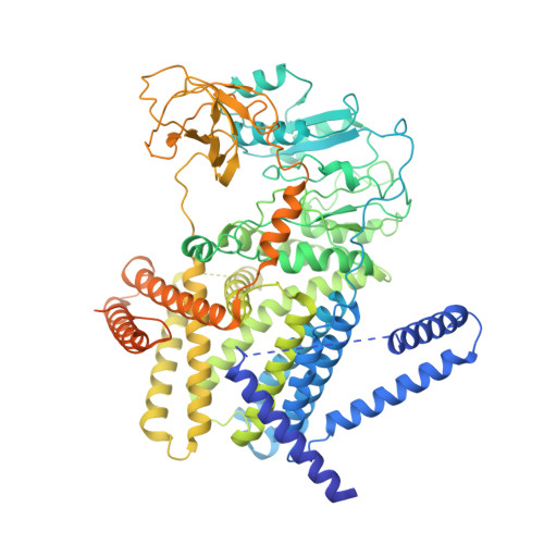
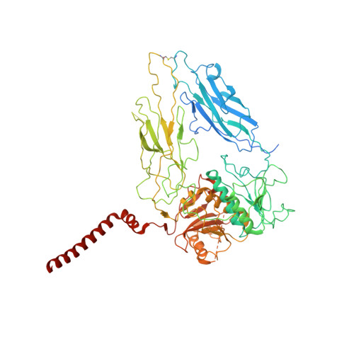
    Cellulose is frequently found in communities of sessile bacteria called biofilms. Escherichia coli and other enterobacteriaceae modify cellulose with phosphoethanolamine (pEtN) to promote host tissue adhesion. The E. coli pEtN cellulose biosynthesis machinery contains the catalytic BcsA-B complex that synthesizes and secretes cellulose, in addition to five other subunits. The membrane-anchored periplasmic BcsG subunit catalyzes pEtN modification. Here we present the structure of the roughly 1 MDa E. coli Bcs complex, consisting of one BcsA enzyme associated with six copies of BcsB, determined by single-particle cryo-electron microscopy. BcsB homo-oligomerizes primarily through interactions between its carbohydrate-binding domains as well as intermolecular beta-sheet formation. The BcsB hexamer creates a half spiral whose open side accommodates two BcsG subunits, directly adjacent to BcsA's periplasmic channel exit. The cytosolic BcsE and BcsQ subunits associate with BcsA's regulatory PilZ domain. The macrocomplex is a fascinating example of cellulose synthase specification.


  • Organizational Affiliation

    Department of Molecular Physiology and Biological Physics, University of Virginia School of Medicine, Charlottesville, VA, USA.


Macromolecules
Find similar proteins by:  (by identity cutoff)  |  3D Structure
Entity ID: 1
MoleculeChains Sequence LengthOrganismDetailsImage
Cellulose synthase catalytic subunit [UDP-forming]886Escherichia coli K-12Mutation(s): 0 
Gene Names: bcsAyhjOyhjPb3533JW5665
EC: 2.4.1.12
Membrane Entity: Yes 
UniProt
Find proteins for P37653 (Escherichia coli (strain K12))
Explore P37653 
Go to UniProtKB:  P37653
Entity Groups  
Sequence Clusters30% Identity50% Identity70% Identity90% Identity95% Identity100% Identity
UniProt GroupP37653
Sequence Annotations
Expand
  • Reference Sequence
Find similar proteins by:  (by identity cutoff)  |  3D Structure
Entity ID: 2
MoleculeChains Sequence LengthOrganismDetailsImage
Cyclic di-GMP-binding protein779Escherichia coli K-12Mutation(s): 0 
Gene Names: bcsByhjNb3532JW3500
Membrane Entity: Yes 
UniProt
Find proteins for P37652 (Escherichia coli (strain K12))
Explore P37652 
Go to UniProtKB:  P37652
Entity Groups  
Sequence Clusters30% Identity50% Identity70% Identity90% Identity95% Identity100% Identity
UniProt GroupP37652
Sequence Annotations
Expand
  • Reference Sequence
Experimental Data & Validation

Experimental Data

  • Method: ELECTRON MICROSCOPY
  • Resolution: 4.20 Å
  • Aggregation State: PARTICLE 
  • Reconstruction Method: SINGLE PARTICLE 
EM Software:
TaskSoftware PackageVersion
MODEL REFINEMENTPHENIX1.18

Structure Validation

View Full Validation Report


Entry History & Funding Information

Deposition Data


Funding OrganizationLocationGrant Number
National Institutes of Health/National Institute of General Medical Sciences (NIH/NIGMS)United States5R01GM101001
National Institutes of Health/National Institute of General Medical Sciences (NIH/NIGMS)United StatesU24-GM116790
National Institutes of Health/National Institute of General Medical Sciences (NIH/NIGMS)United States1F32GM126647-01

Revision History  (Full details and data files)

  • Version 1.0: 2021-03-24
    Type: Initial release