7KCW

Crystal structure of S. aureus penicillin-binding protein 4 (PBP4) mutant (R200L) in complex with nafcillin


Experimental Data Snapshot

  • Method: X-RAY DIFFRACTION
  • Resolution: 1.73 Å
  • R-Value Free: 0.212 
  • R-Value Work: 0.177 
  • R-Value Observed: 0.178 

wwPDB Validation   3D Report Full Report


Ligand Structure Quality Assessment 


This is version 1.3 of the entry. See complete history


Literature

PBP4-mediated beta-lactam resistance among clinical strains of Staphylococcus aureus.

Satishkumar, N.Alexander, J.A.N.Poon, R.Buggeln, E.Argudin, M.A.Strynadka, N.C.J.Chatterjee, S.S.

(2021) J Antimicrob Chemother 76: 2268-2272

  • DOI: https://doi.org/10.1093/jac/dkab201
  • Primary Citation of Related Structures:  
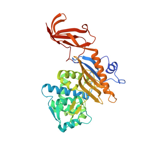
    7KCV, 7KCW, 7KCX, 7KCY

  • PubMed Abstract: 

    PBP4, a low-molecular-weight PBP in Staphylococcus aureus, is not considered to be a classical mediator of β-lactam resistance. Previous studies carried out by our group with laboratory strains of S. aureus demonstrated the ability of PBP4 to produce β-lactam resistance through mutations associated with the pbp4 promoter and/or gene. Recent studies of β-lactam-resistant clinical isolates of S. aureus have reported similar mutations associated with pbp4.


  • Organizational Affiliation

    Department of Microbial Pathogenesis, School of Dentistry, University of Maryland, Baltimore, MD, USA.


Macromolecules
Find similar proteins by:  (by identity cutoff)  |  3D Structure
Entity ID: 1
MoleculeChains Sequence LengthOrganismDetailsImage
Penicillin-binding protein 4367Staphylococcus aureus subsp. aureus COLMutation(s): 1 
Gene Names: pbp4SACOL0699
UniProt
Find proteins for A0A0H2WY27 (Staphylococcus aureus (strain COL))
Explore A0A0H2WY27 
Go to UniProtKB:  A0A0H2WY27
Entity Groups  
Sequence Clusters30% Identity50% Identity70% Identity90% Identity95% Identity100% Identity
UniProt GroupA0A0H2WY27
Sequence Annotations
Expand
  • Reference Sequence
Experimental Data & Validation

Experimental Data

  • Method: X-RAY DIFFRACTION
  • Resolution: 1.73 Å
  • R-Value Free: 0.212 
  • R-Value Work: 0.177 
  • R-Value Observed: 0.178 
  • Space Group: P 21 21 21
Unit Cell:
Length ( Å )Angle ( ˚ )
a = 50.15α = 90
b = 82.87β = 90
c = 104.63γ = 90
Software Package:
Software NamePurpose
PHENIXrefinement
Aimlessdata scaling
PHASERphasing
PDB_EXTRACTdata extraction
XDSdata reduction

Structure Validation

View Full Validation Report


Ligand Structure Quality Assessment 


Entry History & Funding Information

Deposition Data


Funding OrganizationLocationGrant Number
Canadian Institutes of Health Research (CIHR)Canada--

Revision History  (Full details and data files)

  • Version 1.0: 2021-06-30
    Type: Initial release
  • Version 1.1: 2021-08-18
    Changes: Database references
  • Version 1.2: 2021-09-29
    Changes: Database references
  • Version 1.3: 2023-10-18
    Changes: Data collection, Refinement description