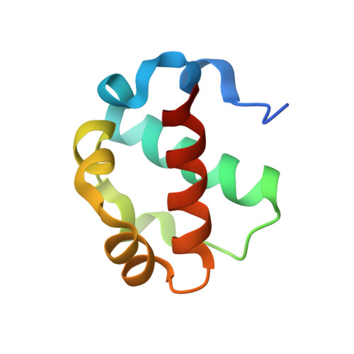
Biophysical characterization of the ETV6 PNT domain polymerization interfaces.
Gerak, C.A.N., Cho, S.Y., Kolesnikov, M., Okon, M., Murphy, M.E.P., Sessions, R.B., Roberge, M., McIntosh, L.P.(2021) J Biol Chem 296: 100284-100284
- PubMed: 33450226
- DOI: https://doi.org/10.1016/j.jbc.2021.100284
- Primary Citation of Related Structures:
7JU2 - PubMed Abstract:
ETV6 is an E26 transformation specific family transcriptional repressor that self-associates by its PNT domain to facilitate cooperative DNA binding. Chromosomal translocations frequently generate constitutively active oncoproteins with the ETV6 PNT domain fused to the kinase domain of one of many protein tyrosine kinases. Although an attractive target for therapeutic intervention, the propensity of the ETV6 PNT domain to polymerize via the tight head-to-tail association of two relatively flat interfaces makes it challenging to identify suitable small molecule inhibitors of this protein-protein interaction. Herein, we provide a comprehensive biophysical characterization of the ETV6 PNT domain interaction interfaces to aid future drug discovery efforts and help define the mechanisms by which its self-association mediates transcriptional repression. Using NMR spectroscopy, X-ray crystallography, and molecular dynamics simulations, along with amide hydrogen exchange measurements, we demonstrate that monomeric PNT domain variants adopt very stable helical bundle folds that do not change in conformation upon self-association into heterodimer models of the ETV6 polymer. Surface plasmon resonance-monitored alanine scanning mutagenesis studies identified hot spot regions within the self-association interfaces. These regions include both central hydrophobic residues and flanking salt-bridging residues. Collectively, these studies indicate that small molecules targeted to these hydrophobic or charged regions within the relatively rigid interfaces could potentially serve as orthosteric inhibitors of ETV6 PNT domain polymerization.
Organizational Affiliation:
Department of Biochemistry and Molecular Biology, University of British Columbia, Vancouver, British Columbia, Canada.