Probing the catalytic mechanism of sulfite reductase by X-ray crystallography: structures of the Escherichia coli hemoprotein in complex with substrates, inhibitors, intermediates, and products.
Crane, B.R., Siegel, L.M., Getzoff, E.D.(1997) Biochemistry 36: 12120-12137
- PubMed: 9315849
- DOI: https://doi.org/10.1021/bi971066i
- Primary Citation of Related Structures:
2GEP, 3GEO, 4GEP, 5GEP, 6GEP, 7GEP, 8GEP - PubMed Abstract:
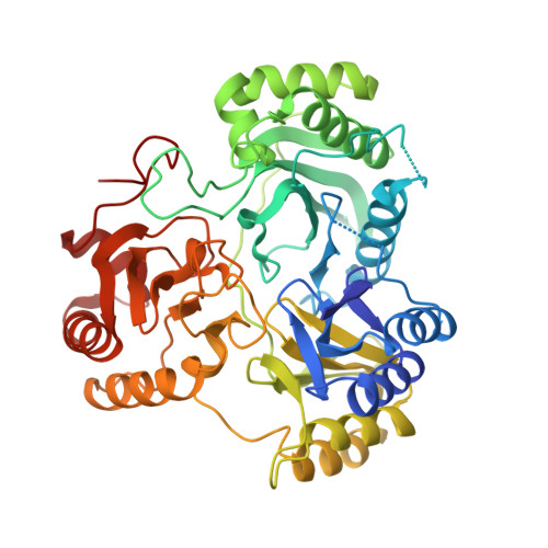
To further understand the six-electron reductions of sulfite and nitrite catalyzed by the Escherichia coli sulfite reductase hemoprotein (SiRHP), we have determined crystallographic structures of the enzyme in complex with the inhibitors phosphate, carbon monoxide, and cyanide, the substrates sulfite and nitrite, the intermediate nitric oxide, the product sulfide (or, most likely, an oxidized derivative thereof), and an oxidized nitrogen species (probably nitrate). A hydrogen-bonded cage of ligand-binding arginine and lysine side chains, ordered water molecules, and siroheme carboxylates provides preferred locations for recognizing the common functional groups of these ligands and accommodates their varied sizes, shapes, and charged without requiring substantial structural changes. The coordination geometries presented here suggest that the successively deoxygenated sulfur and nitrogen species produced during catalysis need not alter their orientation in the active site to adopt new stable coordination states. Strong pi-acid ligands decrease the bond length between the siroheme and the proximal cysteine thiolate shared with the iron-sulfur cluster, emphasizing the ability of the coupled cofactors to promote electron tranfer into substrate. On binding the siroheme, the substrate sulfite provides an oxygen atom in a unique location of the binding site compared to all other ligands studied, induces a spin transition in the siroheme iron, flips an active-site arginine, and orders surrounding active-center loops. The loop that coalesces over the active center shields the positively charged ligand-coordinating residues from solvent, enhancing their ability to polarize the substrate. Hydrogen bonds supplied by active-site arginine and lysine residues facilitate charge transfer into the substrate from the electron-rich cofactors, activate S-O bonds for reductive cleavage, and provide potential proton sources for the formation of favorable aquo leaving groups on the substrate. Strong interactions between sulfite and ordered water molecules also implicate solvent as a source of protons for generating product water. From the structures reported here, we propose a series of key structural states of ligated SiRHP in the catalytic reduction of sulfite to sulfide.
Organizational Affiliation:
Department of Molecular Biology, The Scripps Research Institute, La Jolla, California 92037, USA.