7EEQ

Cyanophage Pam1 tail machine


Experimental Data Snapshot

  • Method: ELECTRON MICROSCOPY
  • Resolution: 3.96 Å
  • Aggregation State: PARTICLE 
  • Reconstruction Method: SINGLE PARTICLE 

wwPDB Validation   3D Report Full Report


This is version 1.1 of the entry. See complete history


Literature

Structure and assembly pattern of a freshwater short-tailed cyanophage Pam1.

Zhang, J.T.Yang, F.Du, K.Li, W.F.Chen, Y.Jiang, Y.L.Li, Q.Zhou, C.Z.

(2022) Structure 30: 240-251.e4

  • DOI: https://doi.org/10.1016/j.str.2021.10.004
  • Primary Citation of Related Structures:  
    7EEA, 7EEL, 7EEP, 7EEQ

  • PubMed Abstract: 

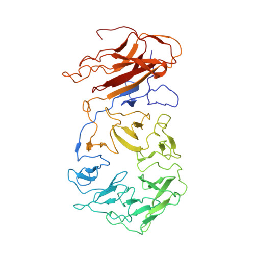
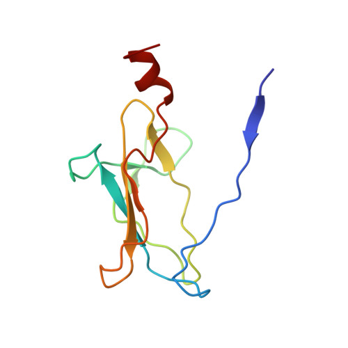
    Despite previous structural analyses of bacteriophages, quite little is known about the structures and assembly patterns of cyanophages. Using cryo-EM combined with crystallography, we solve the near-atomic-resolution structure of a freshwater short-tailed cyanophage, Pam1, which comprises a 400-Å-long tail and an icosahedral capsid of 650 Å in diameter. The outer capsid surface is reinforced by trimeric cement proteins with a β-sandwich fold, which structurally resemble the distal motif of Pam1's tailspike, suggesting its potential role in host recognition. At the portal vertex, the dodecameric portal and connected adaptor, followed by a hexameric needle head, form a DNA ejection channel, which is sealed by a trimeric needle. Moreover, we identify a right-handed rifling pattern that might help DNA to revolve along the wall of the ejection channel. Our study reveals the precise assembly pattern of a cyanophage and lays the foundation to support its practical biotechnological and environmental applications.


  • Organizational Affiliation

    Hefei National Laboratory for Physical Sciences at the Microscale and School of Life Sciences, University of Science and Technology of China, Hefei, Anhui 230026, China.


Macromolecules
Find similar proteins by:  (by identity cutoff)  |  3D Structure
Entity ID: 1
MoleculeChains Sequence LengthOrganismDetailsImage
Needle head proteins442unidentifiedMutation(s): 0 
Entity Groups  
Sequence Clusters30% Identity50% Identity70% Identity90% Identity95% Identity100% Identity
Sequence Annotations
Expand
  • Reference Sequence
Find similar proteins by:  (by identity cutoff)  |  3D Structure
Entity ID: 2
MoleculeChains Sequence LengthOrganismDetailsImage
Tailspike head-binding domain102unidentifiedMutation(s): 0 
Entity Groups  
Sequence Clusters30% Identity50% Identity70% Identity90% Identity95% Identity100% Identity
Sequence Annotations
Expand
  • Reference Sequence
Experimental Data & Validation

Experimental Data

  • Method: ELECTRON MICROSCOPY
  • Resolution: 3.96 Å
  • Aggregation State: PARTICLE 
  • Reconstruction Method: SINGLE PARTICLE 
EM Software:
TaskSoftware PackageVersion
RECONSTRUCTIONRELION3.1

Structure Validation

View Full Validation Report


Entry History & Funding Information

Deposition Data


Funding OrganizationLocationGrant Number
Ministry of Science and Technology (MoST, China)China2018YFA0903100

Revision History  (Full details and data files)

  • Version 1.0: 2021-10-20
    Type: Initial release
  • Version 1.1: 2022-05-04
    Changes: Database references