7AE9

Crystal structure of mono-AMPylated HEPN(R46E) toxin in complex with MNT antitoxin


Experimental Data Snapshot

  • Method: X-RAY DIFFRACTION
  • Resolution: 2.90 Å
  • R-Value Free: 0.246 
  • R-Value Work: 0.203 
  • R-Value Observed: 0.207 

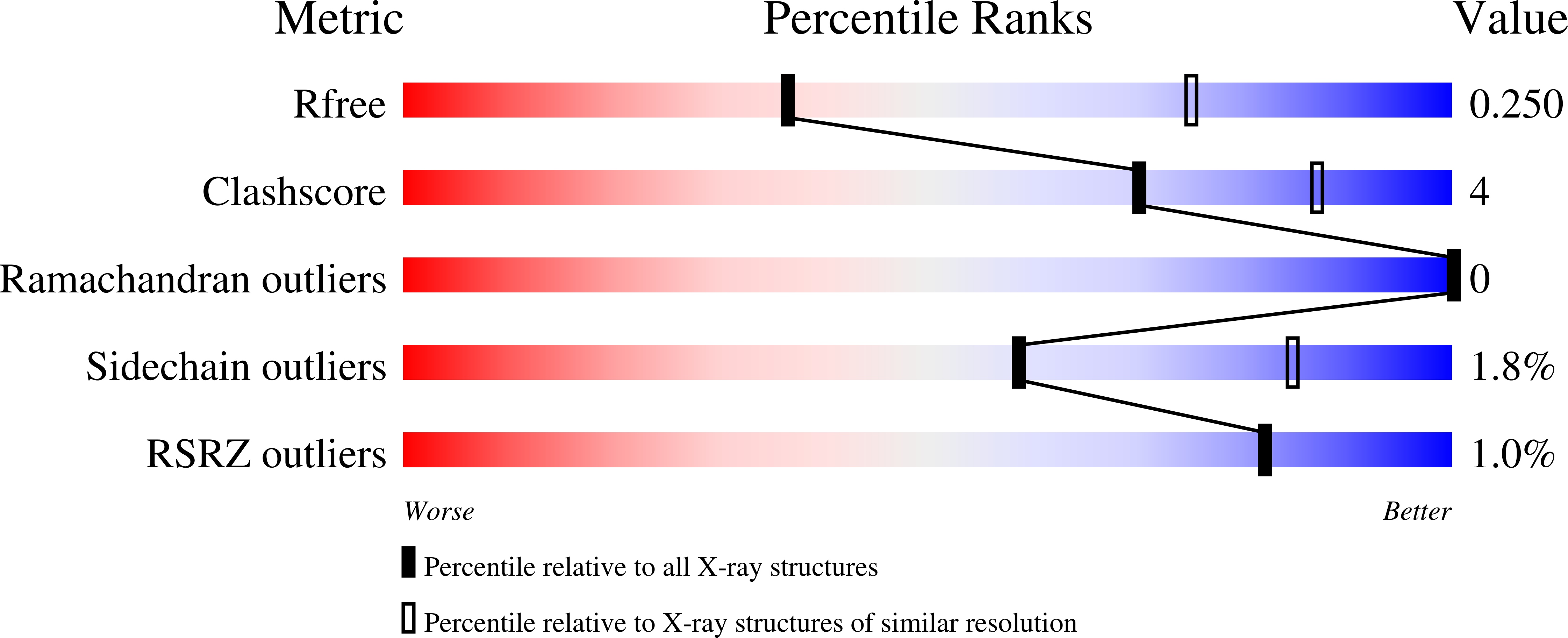
wwPDB Validation   3D Report Full Report


Ligand Structure Quality Assessment 


This is version 1.1 of the entry. See complete history


Literature

HEPN-MNT Toxin-Antitoxin System: The HEPN Ribonuclease Is Neutralized by OligoAMPylation.

Songailiene, I.Juozapaitis, J.Tamulaitiene, G.Ruksenaite, A.Sulcius, S.Sasnauskas, G.Venclovas, C.Siksnys, V.

(2020) Mol Cell 80: 955-970.e7

  • DOI: https://doi.org/10.1016/j.molcel.2020.11.034
  • Primary Citation of Related Structures:  
    7AE2, 7AE6, 7AE8, 7AE9, 7AER

  • PubMed Abstract: 

    Prokaryotic toxin-antitoxin (TA) systems are composed of a toxin capable of interfering with key cellular processes and its neutralizing antidote, the antitoxin. Here, we focus on the HEPN-MNT TA system encoded in the vicinity of a subtype I-D CRISPR-Cas system in the cyanobacterium Aphanizomenon flos-aquae. We show that HEPN acts as a toxic RNase, which cleaves off 4 nt from the 3' end in a subset of tRNAs, thereby interfering with translation. Surprisingly, we find that the MNT (minimal nucleotidyltransferase) antitoxin inhibits HEPN RNase through covalent di-AMPylation (diadenylylation) of a conserved tyrosine residue, Y109, in the active site loop. Furthermore, we present crystallographic snapshots of the di-AMPylation reaction at different stages that explain the mechanism of HEPN RNase inactivation. Finally, we propose that the HEPN-MNT system functions as a cellular ATP sensor that monitors ATP homeostasis and, at low ATP levels, releases active HEPN toxin.


  • Organizational Affiliation

    Institute of Biotechnology, Life Sciences Center, Vilnius University, Sauletekio av. 7, 10257 Vilnius, Lithuania.


Macromolecules
Find similar proteins by:  (by identity cutoff)  |  3D Structure
Entity ID: 1
MoleculeChains Sequence LengthOrganismDetailsImage
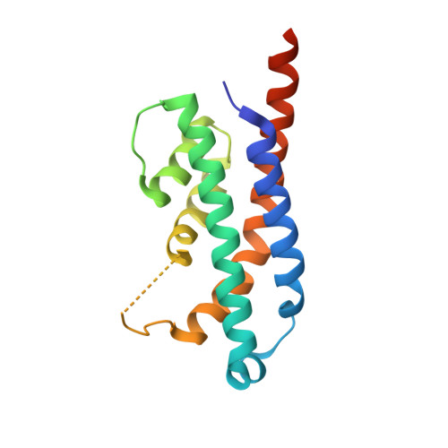
HEPN toxin
A, B, C, D
157Aphanizomenon flos-aquae 2012/KM1/D3Mutation(s): 1 
Gene Names: OA07_26455
UniProt
Find proteins for A0A0B0QJR1 (Aphanizomenon flos-aquae (strain 2012/KM1/D3))
Explore A0A0B0QJR1 
Go to UniProtKB:  A0A0B0QJR1
Entity Groups  
Sequence Clusters30% Identity50% Identity70% Identity90% Identity95% Identity100% Identity
UniProt GroupA0A0B0QJR1
Sequence Annotations
Expand
  • Reference Sequence
Find similar proteins by:  (by identity cutoff)  |  3D Structure
Entity ID: 2
MoleculeChains Sequence LengthOrganismDetailsImage
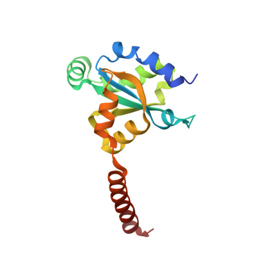
MNT ANTITOXIN
E, F, G, H
150Aphanizomenon flos-aquae 2012/KM1/D3Mutation(s): 0 
Gene Names: OA07_26450
UniProt
Find proteins for A0A0B0QJN8 (Aphanizomenon flos-aquae (strain 2012/KM1/D3))
Explore A0A0B0QJN8 
Go to UniProtKB:  A0A0B0QJN8
Entity Groups  
Sequence Clusters30% Identity50% Identity70% Identity90% Identity95% Identity100% Identity
UniProt GroupA0A0B0QJN8
Sequence Annotations
Expand
  • Reference Sequence
Experimental Data & Validation

Experimental Data

  • Method: X-RAY DIFFRACTION
  • Resolution: 2.90 Å
  • R-Value Free: 0.246 
  • R-Value Work: 0.203 
  • R-Value Observed: 0.207 
  • Space Group: P 21 21 2
Unit Cell:
Length ( Å )Angle ( ˚ )
a = 101.25α = 90
b = 204.543β = 90
c = 73.258γ = 90
Software Package:
Software NamePurpose
PHENIXrefinement
XDSdata reduction
SCALAdata scaling
PHASERphasing
PDB_EXTRACTdata extraction

Structure Validation

View Full Validation Report


Ligand Structure Quality Assessment 


Entry History 

Deposition Data

Revision History  (Full details and data files)

  • Version 1.0: 2020-12-30
    Type: Initial release
  • Version 1.1: 2024-01-31
    Changes: Data collection, Database references, Refinement description