


7VTM
Crystal structure of Glucoside hydrolase family 64 beta-1,3-glucanase complexed with Laminaritetraose
- PDB DOI: https://doi.org/10.2210/pdb7VTM/pdb
- Classification: HYDROLASE
- Organism(s): Streptomyces pratensis
- Expression System: Escherichia coli
- Mutation(s): No
- Deposited: 2021-10-29 Released: 2022-11-02
- Funding Organization(s): National Natural Science Foundation of China (NSFC)
Experimental Data Snapshot
- Method: X-RAY DIFFRACTION
- Resolution: 2.05 Å
- R-Value Free: 0.228
- R-Value Work: 0.211
- R-Value Observed: 0.212
wwPDB Validation 3D Report Full Report

This is version 1.1 of the entry. See complete history.

Macromolecules
Find similar proteins by:
(by identity cutoff) | 3D Structure
Entity ID: 1 | |||||
|---|---|---|---|---|---|
| Molecule | Chains | Sequence Length | Organism | Details | Image |
| beta-1,3-Glucanase | 373 | Streptomyces pratensis | Mutation(s): 0 | 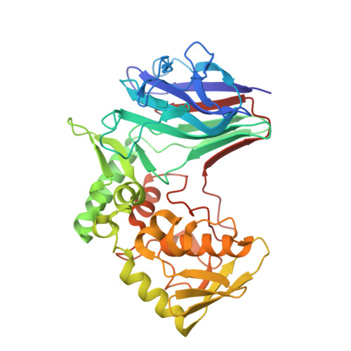 | |
UniProt | |||||
Find proteins for A0A8D3WIQ3 (Streptomyces pratensis (strain ATCC 33331 / IAF-45CD)) Explore A0A8D3WIQ3 Go to UniProtKB: A0A8D3WIQ3 | |||||
Entity Groups | |||||
| Sequence Clusters | 30% Identity50% Identity70% Identity90% Identity95% Identity100% Identity | ||||
| UniProt Group | A0A8D3WIQ3 | ||||
Sequence AnnotationsExpand | |||||
| |||||
Oligosaccharides

Experimental Data & Validation
Experimental Data
- Method: X-RAY DIFFRACTION
- Resolution: 2.05 Å
- R-Value Free: 0.228
- R-Value Work: 0.211
- R-Value Observed: 0.212
- Space Group: P 32 2 1
Unit Cell:
| Length ( Å ) | Angle ( ˚ ) |
|---|---|
| a = 79.486 | α = 90 |
| b = 79.486 | β = 90 |
| c = 156.662 | γ = 120 |
| Software Name | Purpose |
|---|---|
| PHENIX | refinement |
| HKL-3000 | data reduction |
| HKL-3000 | data scaling |
| PHENIX | model building |
| PHENIX | phasing |
Entry History & Funding Information
Deposition Data
- Released Date: 2022-11-02 Deposition Author(s): Jiang, Z.Q., Ma, J.W.
| Funding Organization | Location | Grant Number |
|---|---|---|
| National Natural Science Foundation of China (NSFC) | China | -- |
Revision History (Full details and data files)
- Version 1.0: 2022-11-02
Type: Initial release - Version 1.1: 2023-11-29
Changes: Data collection, Refinement description
