Dimeric architecture of maltodextrin glucosidase (MalZ) provides insights into the substrate recognition and hydrolysis mechanism.
Ahn, W.C., An, Y., Song, K.M., Park, K.H., Lee, S.J., Oh, B.H., Park, J.T., Woo, E.J.(2022) Biochem Biophys Res Commun 586: 49-54
- PubMed: 34826700
- DOI: https://doi.org/10.1016/j.bbrc.2021.11.070
- Primary Citation of Related Structures:
7VT9 - PubMed Abstract:
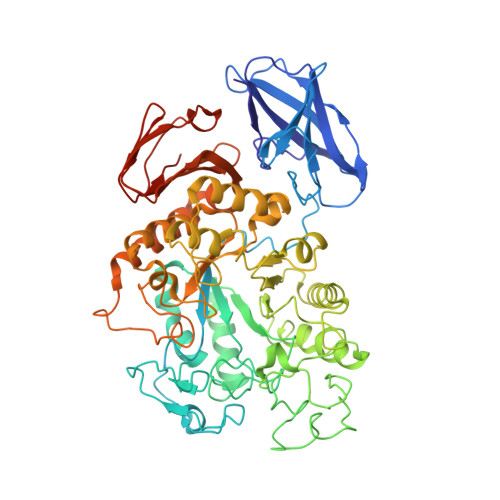
Maltodextrin glucosidase (MalZ) is a key enzyme in the maltose utilization pathway in Escherichia coli that liberates glucose from the reducing end of the short malto-oligosaccharides. Unlike other enzymes in the GH13_21 subfamily, the hydrolytic activity of MalZ is limited to maltodextrin rather than long starch substrates, forming various transglycosylation products in α-1,3, α-1,4 or α-1,6 linkages. The mechanism for the substrate binding and hydrolysis of this enzyme is not well understood yet. Here, we present the dimeric crystal structure of MalZ, with the N-domain generating a unique substrate binding groove. The N-domain bears CBM34 architecture and forms a part of the active site in the catalytic domain of the adjacent molecule. The groove found between the N-domain and catalytic domain from the adjacent molecule, shapes active sites suitable for short malto-oligosaccharides, but hinders long stretches of oligosaccharides. The conserved residue of E44 protrudes at subsite +2, elucidating the hydrolysis pattern of the substrate by the glucose unit from the reducing end. The structural analysis provides a molecular basis for the substrate specificity and the enzymatic property, and has potential industrial application for protein engineering.
Organizational Affiliation:
Department of Biological Science, Korea Advanced Institute of Science and Technology, Daejeon, 34141, Republic of Korea; Disease Target Structure Research Center, Korea Research Institute of Bioscience and Biotechnology, Daejeon, 34141, Republic of Korea.