7UHS

SSX Structure of Metallo Beta-Lactamase L1 with Two Water Molecules in the Active Site


Experimental Data Snapshot

  • Method: X-RAY DIFFRACTION
  • Resolution: 2.20 Å
  • R-Value Free: 0.258 
  • R-Value Work: 0.221 
  • R-Value Observed: 0.223 

wwPDB Validation   3D Report Full Report


This is version 1.2 of the entry. See complete history


Literature

Time-resolved beta-lactam cleavage by L1 metallo-beta-lactamase.

Wilamowski, M.Sherrell, D.A.Kim, Y.Lavens, A.Henning, R.W.Lazarski, K.Shigemoto, A.Endres, M.Maltseva, N.Babnigg, G.Burdette, S.C.Srajer, V.Joachimiak, A.

(2022) Nat Commun 13: 7379-7379

  • DOI: https://doi.org/10.1038/s41467-022-35029-3
  • Primary Citation of Related Structures:  
    7L91, 7UHH, 7UHI, 7UHJ, 7UHK, 7UHL, 7UHM, 7UHN, 7UHO, 7UHP, 7UHQ, 7UHR, 7UHS, 7UHT

  • PubMed Abstract: 

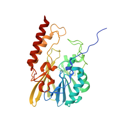
    Serial x-ray crystallography can uncover binding events, and subsequent chemical conversions occurring during enzymatic reaction. Here, we reveal the structure, binding and cleavage of moxalactam antibiotic bound to L1 metallo-β-lactamase (MBL) from Stenotrophomonas maltophilia. Using time-resolved serial synchrotron crystallography, we show the time course of β-lactam hydrolysis and determine ten snapshots (20, 40, 60, 80, 100, 150, 300, 500, 2000 and 4000 ms) at 2.20 Å resolution. The reaction is initiated by laser pulse releasing Zn 2+ ions from a UV-labile photocage. Two metal ions bind to the active site, followed by binding of moxalactam and the intact β-lactam ring is observed for 100 ms after photolysis. Cleavage of β-lactam is detected at 150 ms and the ligand is significantly displaced. The reaction product adjusts its conformation reaching steady state at 2000 ms corresponding to the relaxed state of the enzyme. Only small changes are observed in the positions of Zn 2+ ions and the active site residues. Mechanistic details captured here can be generalized to other MBLs.


  • Organizational Affiliation

    Center for Structural Genomics of Infectious Diseases, Consortium for Advanced Science and Engineering, University of Chicago, Chicago, IL, 60667, USA.


Macromolecules
Find similar proteins by:  (by identity cutoff)  |  3D Structure
Entity ID: 1
MoleculeChains Sequence LengthOrganismDetailsImage
Putative metallo-beta-lactamase l1 (Beta-lactamase type ii) (Ec 3.5.2.6) (Penicillinase)275Stenotrophomonas maltophilia K279aMutation(s): 0 
Gene Names: Smlt2667
EC: 3.5.2.6
UniProt
Find proteins for B2FTM1 (Stenotrophomonas maltophilia (strain K279a))
Explore B2FTM1 
Go to UniProtKB:  B2FTM1
Entity Groups  
Sequence Clusters30% Identity50% Identity70% Identity90% Identity95% Identity100% Identity
UniProt GroupB2FTM1
Sequence Annotations
Expand
  • Reference Sequence
Experimental Data & Validation

Experimental Data

  • Method: X-RAY DIFFRACTION
  • Resolution: 2.20 Å
  • R-Value Free: 0.258 
  • R-Value Work: 0.221 
  • R-Value Observed: 0.223 
  • Space Group: P 64 2 2
Unit Cell:
Length ( Å )Angle ( ˚ )
a = 105.951α = 90
b = 105.951β = 90
c = 99.886γ = 120
Software Package:
Software NamePurpose
PHENIXrefinement
MOLREPphasing
cctbx.primedata scaling
DIALSdata reduction

Structure Validation

View Full Validation Report


Entry History & Funding Information

Deposition Data


Funding OrganizationLocationGrant Number
National Institutes of Health/National Institute Of Allergy and Infectious Diseases (NIH/NIAID)United States--

Revision History  (Full details and data files)

  • Version 1.0: 2022-04-13
    Type: Initial release
  • Version 1.1: 2023-02-08
    Changes: Database references
  • Version 1.2: 2023-10-25
    Changes: Data collection, Refinement description