7SDC

Structure of the SARS-CoV-2 main protease in complex with inhibitor MI-09


Experimental Data Snapshot

  • Method: X-RAY DIFFRACTION
  • Resolution: 1.85 Å
  • R-Value Free: 0.300 
  • R-Value Work: 0.276 
  • R-Value Observed: 0.277 

wwPDB Validation   3D Report Full Report


Ligand Structure Quality Assessment 


This is version 1.2 of the entry. See complete history


Literature

A Systematic Survey of Reversibly Covalent Dipeptidyl Inhibitors of the SARS-CoV-2 Main Protease.

Geng, Z.Z.Atla, S.Shaabani, N.Vulupala, V.Yang, K.S.Alugubelli, Y.R.Khatua, K.Chen, P.H.Xiao, J.Blankenship, L.R.Ma, X.R.Vatansever, E.C.Cho, C.D.Ma, Y.Allen, R.Ji, H.Xu, S.Liu, W.R.

(2023) J Med Chem 66: 11040-11055

  • DOI: https://doi.org/10.1021/acs.jmedchem.3c00221
  • Primary Citation of Related Structures:  
    7SD9, 7SDA, 7SDC

  • PubMed Abstract: 

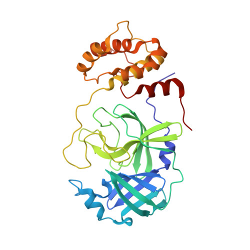
    SARS-CoV-2, the COVID-19 pathogen, relies on its main protease (M Pro ) for replication and pathogenesis. M Pro is a demonstrated target for the development of antivirals for SARS-CoV-2. Past studies have systematically explored tripeptidyl inhibitors such as nirmatrelvir as M Pro inhibitors. However, dipeptidyl inhibitors especially those with a spiro residue at their P2 position have not been systematically investigated. In this work, we synthesized about 30 dipeptidyl M Pro inhibitors and characterized them on enzymatic inhibition potency, structures of their complexes with M Pro , cellular M Pro inhibition potency, antiviral potency, cytotoxicity, and in vitro metabolic stability. Our results indicated that M Pro has a flexible S2 pocket to accommodate inhibitors with a large P2 residue and revealed that dipeptidyl inhibitors with a large P2 spiro residue such as ( S )-2-azaspiro [4,4]nonane-3-carboxylate and ( S )-2-azaspiro[4,5]decane-3-carboxylate have favorable characteristics. One compound, MPI60, containing a P2 ( S )-2-azaspiro[4,4]nonane-3-carboxylate displayed high antiviral potency, low cellular cytotoxicity, and high in vitro metabolic stability.


  • Organizational Affiliation

    Department of Chemistry, Texas A&M Drug Discovery Laboratory, Texas A&M University, College Station, Texas 77843, United States.


Macromolecules
Find similar proteins by:  (by identity cutoff)  |  3D Structure
Entity ID: 1
MoleculeChains Sequence LengthOrganismDetailsImage
3C-like proteinase306Severe acute respiratory syndrome coronavirus 2Mutation(s): 0 
Gene Names: rep1a-1b
UniProt
Find proteins for P0DTD1 (Severe acute respiratory syndrome coronavirus 2)
Explore P0DTD1 
Go to UniProtKB:  P0DTD1
Entity Groups  
Sequence Clusters30% Identity50% Identity70% Identity90% Identity95% Identity100% Identity
UniProt GroupP0DTD1
Sequence Annotations
Expand
  • Reference Sequence
Small Molecules
Ligands 1 Unique
IDChains Name / Formula / InChI Key2D Diagram3D Interactions
I80 (Subject of Investigation/LOI)
Query on I80

Download Ideal Coordinates CCD File 
B [auth A](1R,2S,5S)-N-{(2S)-1-hydroxy-3-[(3S)-2-oxopyrrolidin-3-yl]propan-2-yl}-6,6-dimethyl-3-{[4-(trifluoromethoxy)phenoxy]acetyl}-3-azabicyclo[3.1.0]hexane-2-carboxamide
C24 H30 F3 N3 O6
OINAVIUBZFOBLX-VMBFOHBNSA-N
Experimental Data & Validation

Experimental Data

  • Method: X-RAY DIFFRACTION
  • Resolution: 1.85 Å
  • R-Value Free: 0.300 
  • R-Value Work: 0.276 
  • R-Value Observed: 0.277 
  • Space Group: I 1 2 1
Unit Cell:
Length ( Å )Angle ( ˚ )
a = 54.365α = 90
b = 80.987β = 97.29
c = 87.812γ = 90
Software Package:
Software NamePurpose
PHENIXrefinement
PDB_EXTRACTdata extraction
PROTEUM PLUSdata reduction
Aimlessdata scaling
PHENIXphasing

Structure Validation

View Full Validation Report


Ligand Structure Quality Assessment 


Entry History & Funding Information

Deposition Data


Funding OrganizationLocationGrant Number
Welch FoundationUnited States--

Revision History  (Full details and data files)

  • Version 1.0: 2022-11-09
    Type: Initial release
  • Version 1.1: 2023-10-18
    Changes: Data collection, Refinement description
  • Version 1.2: 2024-03-20
    Changes: Database references