7S4S

Crystal Structure of SARS-CoV-2 S receptor-binding domain (RBD) in complex CoV11 Fab


Experimental Data Snapshot

  • Method: X-RAY DIFFRACTION
  • Resolution: 2.05 Å
  • R-Value Free: 0.198 
  • R-Value Work: 0.166 
  • R-Value Observed: 0.167 

wwPDB Validation   3D Report Full Report


Ligand Structure Quality Assessment 


This is version 1.2 of the entry. See complete history


Literature

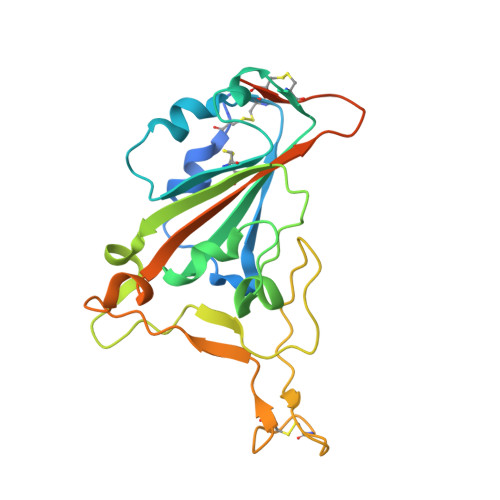
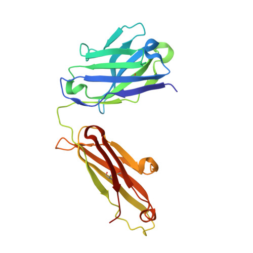
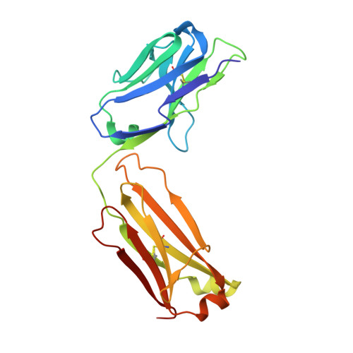
The molecular basis of the neutralization breadth of the RBD-specific antibody CoV11.

Tolbert, W.D.Chen, Y.Sun, L.Benlarbi, M.Ding, S.Manickam, R.Pangaro, E.Nguyen, D.N.Gottumukkala, S.Cote, M.Gonzalez, F.J.Finzi, A.Tehrani, Z.R.Sajadi, M.M.Pazgier, M.

(2023) Front Immunol 14: 1178355-1178355

  • DOI: https://doi.org/10.3389/fimmu.2023.1178355
  • Primary Citation of Related Structures:  
    7S4S, 7URQ, 7URS

  • PubMed Abstract: 

    SARS-CoV-2, the virus behind the COVID-19 pandemic, has changed over time to the extent that the current virus is substantially different from what originally led to the pandemic in 2019-2020. Viral variants have modified the severity and transmissibility of the disease and continue do so. How much of this change is due to viral fitness versus a response to immune pressure is hard to define. One class of antibodies that continues to afford some level of protection from emerging variants are those that closely overlap the binding site for angiotensin-converting enzyme 2 (ACE2) on the receptor binding domain (RBD). Some members of this class that were identified early in the course of the pandemic arose from the V H 3-53 germline gene (IGHV3-53*01) and had short heavy chain complementarity-determining region 3s (CDR H3s). Here, we describe the molecular basis of the SARS-CoV-2 RBD recognition by the anti-RBD monoclonal antibody CoV11 isolated early in the COVID-19 pandemic and show how its unique mode of binding the RBD determines its neutralization breadth. CoV11 utilizes a heavy chain V H 3-53 and a light chain V K 3-20 germline sequence to bind to the RBD. Two of CoV11's four heavy chain changes from the V H 3-53 germline sequence, T h r F W R H 1 28 to Ile and S e r C D R H 1 31 to Arg, and some unique features in its CDR H3 increase its affinity to the RBD, while the four light chain changes from the V K 3-20 germline sequence sit outside of the RBD binding site. Antibodies of this type can retain significant affinity and neutralization potency against variants of concern (VOCs) that have diverged significantly from original virus lineage such as the prevalent omicron variant. We also discuss the mechanism by which V H 3-53 encoded antibodies recognize spike antigen and show how minimal changes to their sequence, their choice of light chain, and their mode of binding influence their affinity and impact their neutralization breadth.


  • Organizational Affiliation

    Infectious Disease Division, Department of Medicine of Uniformed Services University of the Health Sciences, Bethesda, MD, United States.


Macromolecules
Find similar proteins by:  (by identity cutoff)  |  3D Structure
Entity ID: 1
MoleculeChains Sequence LengthOrganismDetailsImage
Spike protein S1231Severe acute respiratory syndrome coronavirus 2Mutation(s): 0 
Gene Names: S2
UniProt
Find proteins for P0DTC2 (Severe acute respiratory syndrome coronavirus 2)
Explore P0DTC2 
Go to UniProtKB:  P0DTC2
Entity Groups  
Sequence Clusters30% Identity50% Identity70% Identity90% Identity95% Identity100% Identity
UniProt GroupP0DTC2
Sequence Annotations
Expand
  • Reference Sequence
Find similar proteins by:  (by identity cutoff)  |  3D Structure
Entity ID: 2
MoleculeChains Sequence LengthOrganismDetailsImage
CoV11 heavy chainB [auth H]221Homo sapiensMutation(s): 0 
Entity Groups  
Sequence Clusters30% Identity50% Identity70% Identity90% Identity95% Identity100% Identity
Sequence Annotations
Expand
  • Reference Sequence
Find similar proteins by:  (by identity cutoff)  |  3D Structure
Entity ID: 3
MoleculeChains Sequence LengthOrganismDetailsImage
CoV11 light chainC [auth L]216Homo sapiensMutation(s): 0 
Entity Groups  
Sequence Clusters30% Identity50% Identity70% Identity90% Identity95% Identity100% Identity
Sequence Annotations
Expand
  • Reference Sequence
Small Molecules
Ligands 3 Unique
IDChains Name / Formula / InChI Key2D Diagram3D Interactions
NAG
Query on NAG

Download Ideal Coordinates CCD File 
D [auth A]2-acetamido-2-deoxy-beta-D-glucopyranose
C8 H15 N O6
OVRNDRQMDRJTHS-FMDGEEDCSA-N
SO4
Query on SO4

Download Ideal Coordinates CCD File 
E [auth A],
G [auth H],
I [auth L],
J [auth L],
K [auth L]
SULFATE ION
O4 S
QAOWNCQODCNURD-UHFFFAOYSA-L
ACT
Query on ACT

Download Ideal Coordinates CCD File 
F [auth A],
H,
L,
M [auth L]
ACETATE ION
C2 H3 O2
QTBSBXVTEAMEQO-UHFFFAOYSA-M
Experimental Data & Validation

Experimental Data

  • Method: X-RAY DIFFRACTION
  • Resolution: 2.05 Å
  • R-Value Free: 0.198 
  • R-Value Work: 0.166 
  • R-Value Observed: 0.167 
  • Space Group: P 21 21 21
Unit Cell:
Length ( Å )Angle ( ˚ )
a = 55.056α = 90
b = 111.259β = 90
c = 142.948γ = 90
Software Package:
Software NamePurpose
PHENIXrefinement
REFMACrefinement
HKL-3000data reduction
HKL-3000data scaling
PHASERphasing

Structure Validation

View Full Validation Report


Ligand Structure Quality Assessment 


Entry History & Funding Information

Deposition Data


Funding OrganizationLocationGrant Number
National Institutes of Health/National Institute Of Allergy and Infectious Diseases (NIH/NIAID)United StatesR01 AI116274
National Institutes of Health/National Institute Of Allergy and Infectious Diseases (NIH/NIAID)United StatesP01 AI120756
National Institutes of Health/National Institute Of Allergy and Infectious Diseases (NIH/NIAID)United StatesR01 AI129769

Revision History  (Full details and data files)

  • Version 1.0: 2021-09-22
    Type: Initial release
  • Version 1.1: 2023-10-18
    Changes: Data collection, Refinement description
  • Version 1.2: 2023-11-15
    Changes: Database references