7RSM

Crystal structure of pyrrolysyl-tRNA synthetase (N346D/C348S/Y384F) in complex with o-Chlorophenylalanine and AMP-PNP


Experimental Data Snapshot

  • Method: X-RAY DIFFRACTION
  • Resolution: 2.15 Å
  • R-Value Free: 0.309 
  • R-Value Work: 0.247 
  • R-Value Observed: 0.250 

wwPDB Validation   3D Report Full Report


Ligand Structure Quality Assessment 


This is version 2.0 of the entry. See complete history


Literature

A Designed, Highly Efficient Pyrrolysyl-tRNA Synthetase Mutant Binds o-Chlorophenylalanine Using Two Halogen Bonds.

Vatansever, E.C.Yang, K.S.Geng, Z.Z.Qiao, Y.Li, P.Xu, S.Liu, W.R.

(2022) J Mol Biol 434: 167534-167534

  • DOI: https://doi.org/10.1016/j.jmb.2022.167534
  • Primary Citation of Related Structures:  
    7RSM

  • PubMed Abstract: 

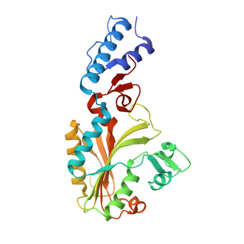
    As one of the most valuable tools for genetic code expansion, pyrrolysyl-tRNA synthetase (PylRS) is structurally related to phenylalanyl-tRNA synthetase (PheRS). By introducing mutations that mimic ligand interactions in PheRS into PylRS, we designed a PylRS mutant. This mutant, designated as oClFRS, recognizes a number of o-substituted phenylalanines for their genetic incorporation at amber codon. Its efficiency in catalyzing genetic incorporation of o-chlorophenylalanine (o-ClF) is better than that for N ε -tert-butyloxycarbonyl-lysine catalyzed by PylRS. The crystal structure of oClFRS bound with o-ClF shows that o-ClF binds deeply into a hydrophobic but catalytically inactive pocket in the active site and involves two halogen bonds to achieve strong interactions. The shift of o-ClF to a catalytically active position in the oClFRS active site will be necessary for its activation. This is the first reported aminoacyl-tRNA synthetase that involves two halogen bonds for ligation recognition and might represent an alternative route to develop aminoacyl-tRNA synthetase mutants that are selective for noncanonical amino acids over native amino acids.


  • Organizational Affiliation

    The Texas A&M Drug Discovery Laboratory, Department of Chemistry, Texas A&M University, College Station, TX 77843, USA.


Macromolecules
Find similar proteins by:  (by identity cutoff)  |  3D Structure
Entity ID: 1
MoleculeChains Sequence LengthOrganismDetailsImage
Pyrrolysine--tRNA ligase
A, B, C, D
267Methanosarcina mazeiMutation(s): 3 
Gene Names: pylSDU43_20175DU67_18120
EC: 6.1.1.26
UniProt
Find proteins for Q8PWY1 (Methanosarcina mazei (strain ATCC BAA-159 / DSM 3647 / Goe1 / Go1 / JCM 11833 / OCM 88))
Explore Q8PWY1 
Go to UniProtKB:  Q8PWY1
Entity Groups  
Sequence Clusters30% Identity50% Identity70% Identity90% Identity95% Identity100% Identity
UniProt GroupQ8PWY1
Sequence Annotations
Expand
  • Reference Sequence
Experimental Data & Validation

Experimental Data

  • Method: X-RAY DIFFRACTION
  • Resolution: 2.15 Å
  • R-Value Free: 0.309 
  • R-Value Work: 0.247 
  • R-Value Observed: 0.250 
  • Space Group: P 1 21 1
Unit Cell:
Length ( Å )Angle ( ˚ )
a = 66.7α = 90
b = 135.2β = 112.54
c = 72.24γ = 90
Software Package:
Software NamePurpose
PHENIXrefinement
PDB_EXTRACTdata extraction
iMOSFLMdata reduction
Aimlessdata scaling
PHENIXphasing

Structure Validation

View Full Validation Report


Ligand Structure Quality Assessment 


Entry History & Funding Information

Deposition Data


Funding OrganizationLocationGrant Number
Welch FoundationUnited StatesA1715

Revision History  (Full details and data files)

  • Version 1.0: 2022-07-20
    Type: Initial release
  • Version 1.1: 2023-10-18
    Changes: Data collection, Refinement description
  • Version 2.0: 2023-11-15
    Changes: Atomic model, Data collection