7R1U

Crystal structure of SARS-CoV-2 nsp10/nsp16 in complex with the WZ16 inhibitor


Experimental Data Snapshot

  • Method: X-RAY DIFFRACTION
  • Resolution: 2.50 Å
  • R-Value Free: 0.252 
  • R-Value Work: 0.232 
  • R-Value Observed: 0.233 

wwPDB Validation   3D Report Full Report


Ligand Structure Quality Assessment 


This is version 1.2 of the entry. See complete history


Literature

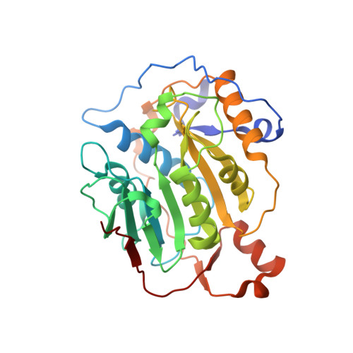
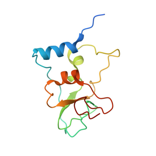
Crystal structure of SARS-CoV-2 nsp10-nsp16 in complex with small molecule inhibitors, SS148 and WZ16.

Klima, M.Khalili Yazdi, A.Li, F.Chau, I.Hajian, T.Bolotokova, A.Kaniskan, H.U.Han, Y.Wang, K.Li, D.Luo, M.Jin, J.Boura, E.Vedadi, M.

(2022) Protein Sci 31: e4395-e4395

  • DOI: https://doi.org/10.1002/pro.4395
  • Primary Citation of Related Structures:  
    7R1T, 7R1U

  • PubMed Abstract: 

    SARS-CoV-2 nsp10-nsp16 complex is a 2'-O-methyltransferase (MTase) involved in viral RNA capping, enabling the virus to evade the immune system in humans. It has been considered a valuable target in the discovery of antiviral therapeutics, as the RNA cap formation is crucial for viral propagation. Through cross-screening of the inhibitors that we previously reported for SARS-CoV-2 nsp14 MTase activity against nsp10-nsp16 complex, we identified two compounds (SS148 and WZ16) that also inhibited nsp16 MTase activity. To further enable the chemical optimization of these two compounds towards more potent and selective dual nsp14/nsp16 MTase inhibitors, we determined the crystal structure of nsp10-nsp16 in complex with each of SS148 and WZ16. As expected, the structures revealed the binding of both compounds to S-adenosyl-L-methionine (SAM) binding pocket of nsp16. However, our structural data along with the biochemical mechanism of action determination revealed an RNA-dependent SAM-competitive pattern of inhibition for WZ16, clearly suggesting that binding of the RNA first may help the binding of some SAM competitive inhibitors. Both compounds also showed some degree of selectivity against human protein MTases, an indication of great potential for chemical optimization towards more potent and selective inhibitors of coronavirus MTases.


  • Organizational Affiliation

    Institute of Organic Chemistry and Biochemistry, Czech Academy of Sciences, Prague 6, Czech Republic.


Macromolecules
Find similar proteins by:  (by identity cutoff)  |  3D Structure
Entity ID: 1
MoleculeChains Sequence LengthOrganismDetailsImage
2'-O-methyltransferase nsp16299Severe acute respiratory syndrome coronavirus 2Mutation(s): 0 
Gene Names: rep1a-1b
EC: 2.1.1.57
UniProt
Find proteins for P0DTD1 (Severe acute respiratory syndrome coronavirus 2)
Explore P0DTD1 
Go to UniProtKB:  P0DTD1
Entity Groups  
Sequence Clusters30% Identity50% Identity70% Identity90% Identity95% Identity100% Identity
UniProt GroupP0DTD1
Sequence Annotations
Expand
  • Reference Sequence
Find similar proteins by:  (by identity cutoff)  |  3D Structure
Entity ID: 2
MoleculeChains Sequence LengthOrganismDetailsImage
Non-structural protein 10123Severe acute respiratory syndrome coronavirus 2Mutation(s): 0 
Gene Names: rep1a-1b
UniProt
Find proteins for P0DTD1 (Severe acute respiratory syndrome coronavirus 2)
Explore P0DTD1 
Go to UniProtKB:  P0DTD1
Entity Groups  
Sequence Clusters30% Identity50% Identity70% Identity90% Identity95% Identity100% Identity
UniProt GroupP0DTD1
Sequence Annotations
Expand
  • Reference Sequence
Small Molecules
Ligands 4 Unique
IDChains Name / Formula / InChI Key2D Diagram3D Interactions
GTA
Query on GTA

Download Ideal Coordinates CCD File 
D [auth A]P1-7-METHYLGUANOSINE-P3-ADENOSINE-5',5'-TRIPHOSPHATE
C21 H30 N10 O17 P3
QQOHNVHGNZYSBP-XPWFQUROSA-O
4IK (Subject of Investigation/LOI)
Query on 4IK

Download Ideal Coordinates CCD File 
C [auth A](2S,5S)-2,6-diamino-5-{[(2R,3S,4R,5R)-5-(6-amino-9H-purin-9-yl)-3,4-dihydroxytetrahydrofuran-2-yl]methyl}hexanoic acid
C16 H25 N7 O5
CPUNUFGYWFHFOF-TVDBPQCTSA-N
MES
Query on MES

Download Ideal Coordinates CCD File 
E [auth A]2-(N-MORPHOLINO)-ETHANESULFONIC ACID
C6 H13 N O4 S
SXGZJKUKBWWHRA-UHFFFAOYSA-N
ZN
Query on ZN

Download Ideal Coordinates CCD File 
F [auth B],
G [auth B]
ZINC ION
Zn
PTFCDOFLOPIGGS-UHFFFAOYSA-N
Experimental Data & Validation

Experimental Data

  • Method: X-RAY DIFFRACTION
  • Resolution: 2.50 Å
  • R-Value Free: 0.252 
  • R-Value Work: 0.232 
  • R-Value Observed: 0.233 
  • Space Group: P 31 2 1
Unit Cell:
Length ( Å )Angle ( ˚ )
a = 168.011α = 90
b = 168.011β = 90
c = 52.085γ = 120
Software Package:
Software NamePurpose
XDSdata reduction
XDSdata scaling
PHASERphasing
Cootmodel building
PHENIXrefinement

Structure Validation

View Full Validation Report


Ligand Structure Quality Assessment 


Entry History & Funding Information

Deposition Data


Funding OrganizationLocationGrant Number
Not funded--

Revision History  (Full details and data files)

  • Version 1.0: 2022-06-29
    Type: Initial release
  • Version 1.1: 2022-09-07
    Changes: Database references
  • Version 1.2: 2024-01-31
    Changes: Data collection, Refinement description